Foot Toe Anatomy
The big toes function is to provide additional leverage to the foot when it pushes off the ground during walking running. Toe anatomy each of your toes is made up of several structures found throughout the body.
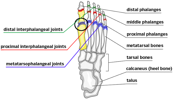 Hallux Rigidus Stiff Big Toe Hss Edu
These make up the toes and broad section of the feet.

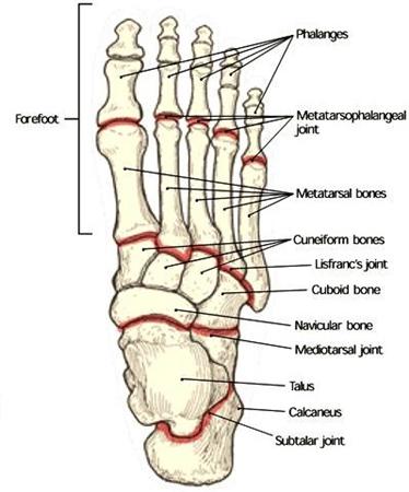
Foot toe anatomy. Hind means posterior so it basically the backward part of the foot. Tarsals five irregularly shaped bones of the midfoot that form the foots arch. At the same time the foot must be strong to support more than 100000 pounds of pressure for every mile walked.
The foot is located after the long shin bones and it starts from the back of your ankle to your toes. An autoimmune form of arthritis that causes inflammation and joint damage. A fungal infection of the feet causing dry flaking red and irritated skin.
The big toe is often affected by gout. The hindfoot midfoot and the forefoot. Each toe is surrounded by skin and present on all five toes is a toenail.
A joint is formed at the junction between two or more bones. The bones of the feet are. The toes are from medial to lateral.
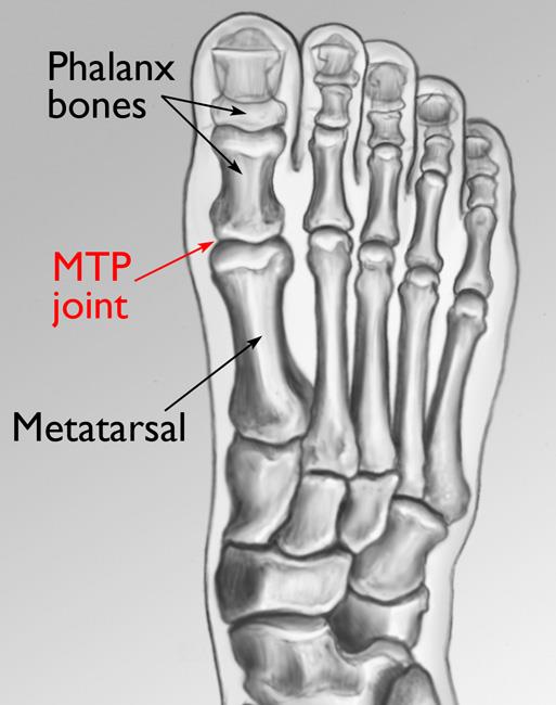
The phalanges which are the bones in your toes. The cuneiform bones the navicularis and the cuboid all of which function to give your foot. The metatarsals which run through the flat part of your foot.
Anatomically the foot is divided into 3 sections. The first toe also known as the hallux big toe great toe thumb toe the innermost toe. Calcaneus the largest bone of the foot which lies beneath the talus to form the heel bone.
The bones of the foot are organized into rows named tarsal bones metatarsal bones and phalanges. Daily washing and keeping the feet dry can prevent athletes foot. The other bones of the foot that create the ankle and connecting bones include.
It is officially known as the hallux. The big toe is one of five digits located on the front of the foot. Talus the bone on top of the foot that forms a joint with the two bones of the lower leg.
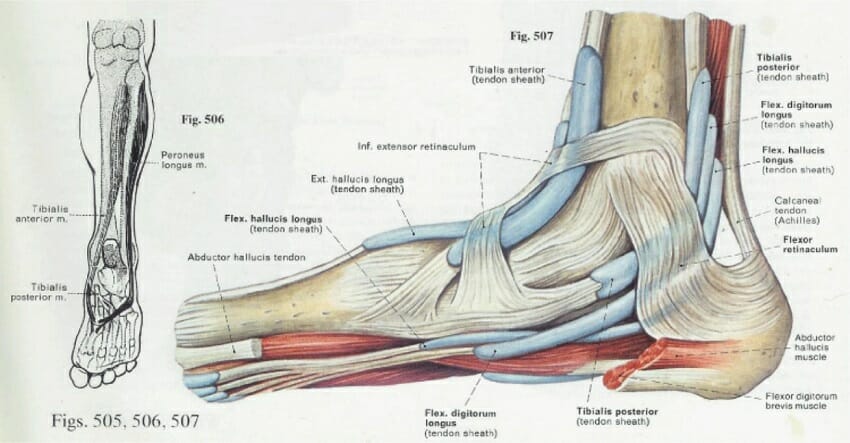
The phalanges are connected to bones of the foot by tendons and muscles allowing them to move and flex. The forefoot midfoot and hindfoot. It is the innermost toe of tetrapods animals that have four limbs and is counted as digit number one.
The proximal phalanx bone of each toe articulates with the metatarsal bone of the foot at the metatarsophalangeal joint. The foot can be divided into three sections. This is the very front part of the foot including the toes or phalanges.
The hindfoot is the posterior part of the foot. They have bones nerves arteries veins tendons and musclesthe bones of the toes like those of the fingers are called phalanges. The talus which is the.
The anatomy of the foot foot structure. The foot is an extremely complex anatomic structure made up of 26 bones and 33 joints that must work together with 19 muscles and 107 ligaments to execute highly precise movements. The calcaneus which is the bone in your heel.
Human Being Anatomy Skeleton Foot Image Visual
 Foot And Leg Anatomy Essential Info For Yoga Teachers
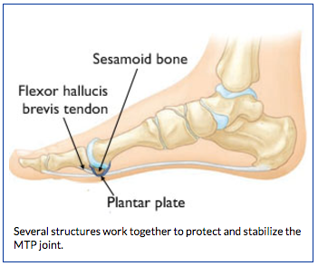 Anatomy Of Turf Toe Bouldercentre For Orthopedics Spine
 Facts About Feet Anatomy Snippets Complete Anatomy
Patient Education Concord Orthopaedics
 Feet Human Anatomy Bones Tendons Ligaments And More
:background_color(FFFFFF):format(jpeg)/images/library/11041/anatomy-ankle-joint_english.jpg) Ankle And Foot Anatomy Bones Joints Muscles Kenhub
Ankle And Foot Anatomy Bones Joints Muscles Kenhub
 Pronation Of The Foot Wikipedia
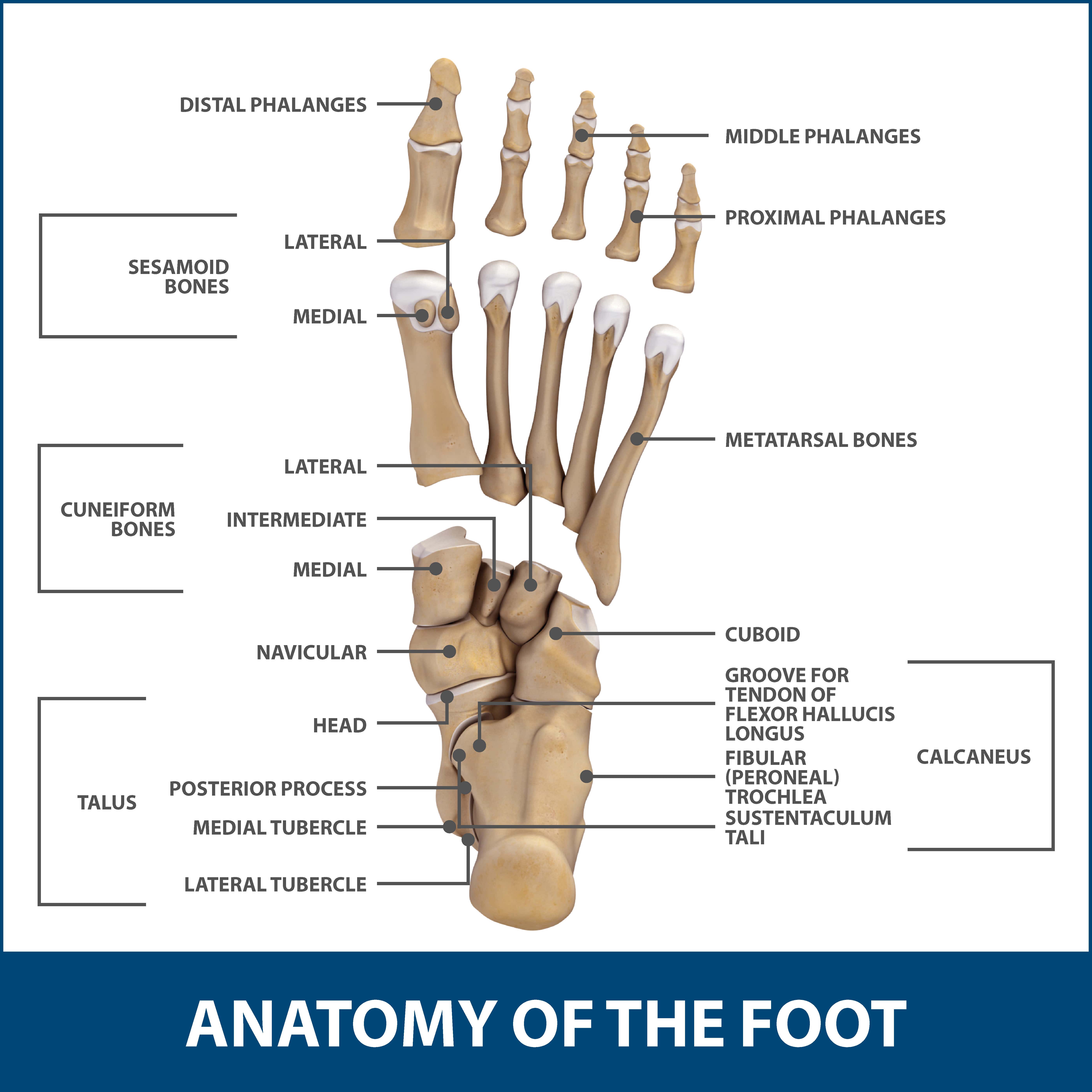 Mallet Hammer Claw Toes Florida Orthopaedic Institute
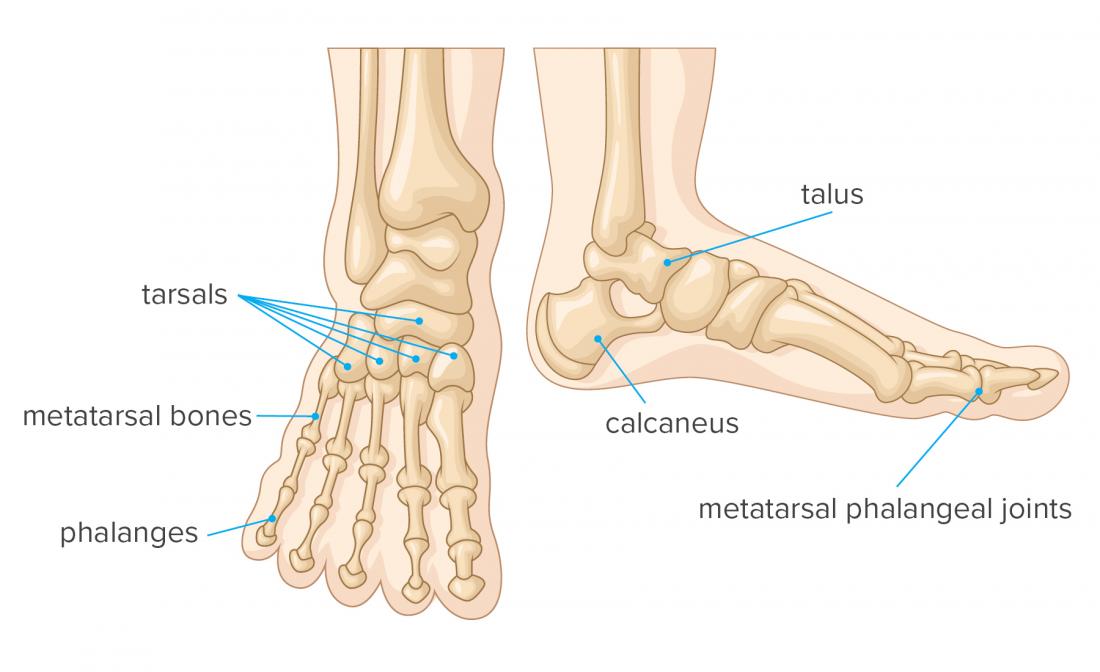 Foot Bones Anatomy Conditions And More
 Foot Anatomy And Biomechanics Foot Ankle Orthobullets
 Abductor Hallucis Muscle Wikipedia
 Foot Anatomy Bones Ligaments Muscles Tendons Arches
1 Ce Credit Radiography Of The Foot E Book And Test
Foot Anatomy Orthopedic Surgery Algonquin Il Barrington
 Skeleton Feet Foot Bones Top View Your Foot Is Quite Easy
 Duke Anatomy Lab 2 Pre Lab Exercise In 2019 Foot Anatomy
 How To Draw The Foot Drawing Feet And The Anatomy Of Them
 Drawing Art Heels Shoes Draw Feet High Heels Anatomy
 Facts About Feet Anatomy Snippets Complete Anatomy
 In 30 Days You Too Can Type And Play The Piano With Your


Belum ada Komentar untuk "Foot Toe Anatomy"
Posting Komentar