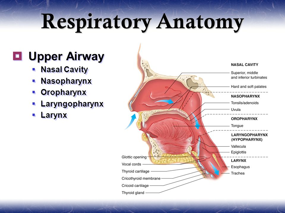
Upper Airway Anatomy
The rear of the laryngopharynx merges with the esophagus to continue the digestive tract. The nose is composed of bone and cartilage which are in turn attached to the facial skeleton.
 Upper Airway Evaluation In Snoring And Obstructive Sleep
Upper airway anatomy and function.

Upper airway anatomy. Functional anatomy of the upper airway. Knowledge of the functional anatomy of the airway in these forms the basis of understanding the pathological conditions that may occur. The airway changes in size shape and position throughout its development from the neonate to the adults.
The upper respiratory and upper digestive tracts diverge after the laryngopharynx. Upper airway anatomy functions warm filter and humidify air nasal cavity and nasopharynx formed by union of facial bones nasal floor towards ear not eye lined with mucous membranes cilia tissues are delicate vascular adenoids lymph tissue filters bacteria commonly infected. The upper airway extends from the mouth to the trachea.
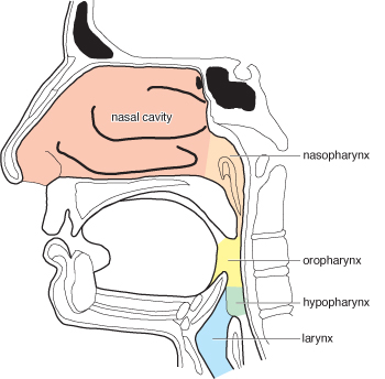
The pharynx is can be divided into the nasopharynx oropharynx and laryngopharynx. The anterior attachments of the middle constrictor are the hyoid bone and the stylohyoid ligament. Anatomy and physiology of the respiratory system duration.
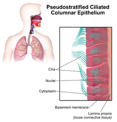
Upper respiratory tract structural and functional anatomy nose and nasal cavity. Unlike the trachea and bronchi the upper airway is a collapsible compliant tube. Lying just after the nostrils are the two nasal cavities lined with mucous membrane and tiny hair like projections called cilia 6.
It evolved as a protective valve mechanism at the upper end of the lower airway necessitated by an unusual crossover between the airway and alimentary canal. The superior constrictor is suspended from the base of the skull the medial pterygoid plate the pterygomandibular raphe the mylohyoid line of the mandible and the lateral tongue. The airway consists of chambers and pipes which conduct air with its 21 oxygen content to the alveoli and carry away the waste carbon dioxide that diffuses from the blood into the alveoli.
The upper airway consists of the pharynx and the nasal cavities. Carbon dioxide co 2 is transferred from returning blood back into gaseous form in the lungs and exhaled through the lower respiratory tract and then the upper to complete the process of breathing. However some authors include the larynx and trachea as well.
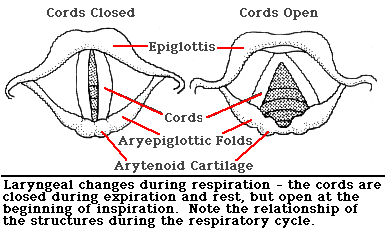
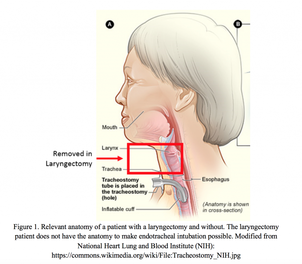
The laryngopharynx is the posteriormost portion of the pharynx reaching from the hyoid to the cricoid cartilage. In this first aid blog post we will look closer at the anatomy of the upper airway. The larynx consists of a framework of cartilages and fibroelastic membranes covered by a sheet of muscles and lined with mucous membrane.
The nostrils the two round or oval holes below the external nose are the primary entrance into the human respiratory system 5.
 Airway Anatomy Nurse Anesthesia Nran 788 With Hadenfeldt
 Rt 127
 Anatomy And Physiology Of Upper Airway Obstruction Neupsy Key
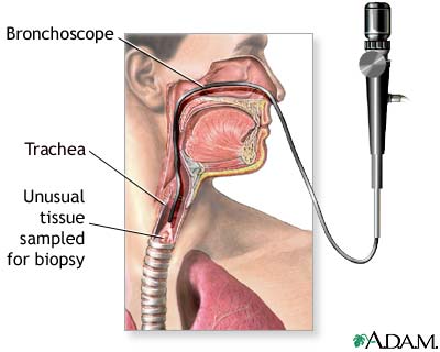 Upper Airway Test Medlineplus Medical Encyclopedia Image
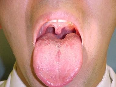
 Healthy Upper Airway Of Child
 Large Airways Radiology Key
 Basic Airway Abdullah Alsakka Em Consultant Objectives
Human Respiratory System Description Parts Function
 Virtual Pediatric Hospital Electricairway Upper Airway
 Pulmonology Ppt Video Online Download

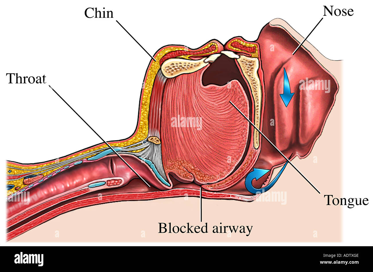 Obstructive Sleep Apnea Blocked Upper Airway Stock Photo

Search Anatomy Upper Airway
 Rt 127

 Rt 127
 Selner S Rhinolaryngoscopy Online Chapter 3 Normal Anatomy
 Acute Upper Airway Obstruction Nejm
 Pdf An Investigation Into The Development Of A Realistic
 Ppt Module A2 Upper Airway Anatomy Physiology
 Respiratory Tract Wikipedia
 Emt Chapter 9 Airway Management At Estrella Mountain
Belum ada Komentar untuk "Upper Airway Anatomy"
Posting Komentar