Anatomy Of Brain Ct
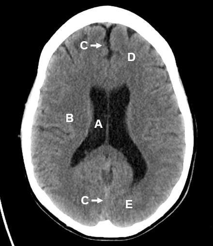
Brain bones of cranium sinuses of the face. Lateral ventricles of normal volume.
 The Radiology Assistant Brain Anatomy
Head ct anatomy normal anatomy 1.

Anatomy of brain ct. Welcome to online mri ct sectional anatomy. Adequate gray matter white matter differentiation. Ct cross sectional anatomy of brain chest abdomen paranasal sinusus neck temporal bone heart slideshare uses cookies to improve functionality and performance and to provide you with relevant advertising.
Anatomy ct axial brain form no 18. Anatomy of the head on a cranial ct scan. Online mri ct sectional anatomy omcsa k anatomy is probably one of the most user friendly and convenient online interface for human anatomy atlas.
Basal subarachnoid cisterns normal configuration. Real time interface human sectional anatomy. Focal abnormalities are not observed in the brain parenchyma.
Brain bones of cranium sinuses of the face. Brainstem and cerebellum without evidence of focal lesions. Brain and face ct.
Ct anatomy of the brain. Anatomy of the head on a cranial ct scan. They lie on the ventricular surface of the hippocampus and become the fimbria of the fornix medially.
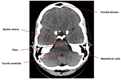
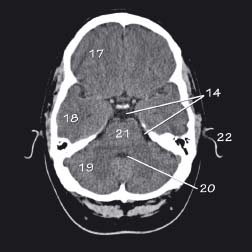
Third and fourth ventricles in midline. 6 frontal bone 27 occipital bone 32 optic nerve 37 basilar artery 40 hemisphere of cerebellum 43 frontal sinus 45 sigmoid sinus 46 internal carotid artery 47 sphenoid bone 49 medulla oblongata 50 external auditory meatus 51 spinal central canal. The anterior part of the head is at the top of the image.
Ct brain image orientation. Amygdala on ct and mr images the amygdala is a large region of gray matter contiguous with the uncus of the medial temporal lobe and the most anterior portion of the hippocampus the pes hippocampi. Ct images of the brain are conventionally viewed from below as if looking up into the top of the head.
This means that the right side of the brain is on the left side of the viewer.
 Basics Of Ct Head
 Ct Brain Hemorrhage Startradiology
 How To Interpret An Unenhanced Ct Brain Scan Part 1 Basic
 Brain Imaging
 Mri Ct Brain For Lobar Anatomy
![]() Medical Imaging And Radiological Anatomy X Ray Ct Mri
Medical Imaging And Radiological Anatomy X Ray Ct Mri
 Ct Medulla
 Ct Anatomy Of Skull
 Brain Imaging
 Brain And Face Ct Interactive Anatomy Atlas
 Brain And Face Ct Interactive Anatomy Atlas
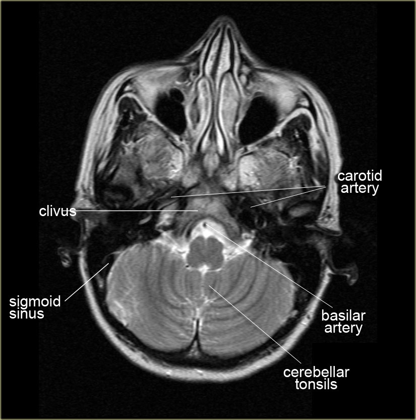
 The Radiology Assistant Brain Anatomy
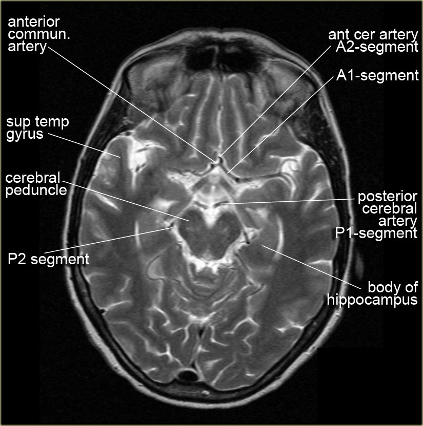
 Mri Anatomy Free Mri Axial Brain Anatomy
 Normal Ct Brain
 How To Interpret An Unenhanced Ct Brain Scan Part 1 Basic
 Normal Brain Ct 2 Month Old Radiology Case Radiopaedia Org
 Mri Anatomy Free Mri Axial Brain Anatomy
 Brain And Face Ct Interactive Anatomy Atlas
 Introduction To Ct Head Approach And Principles
 Computed Tomography Of The Head Wikipedia
 How To Read A Head Ct Emergency Medicine Newyork
 How To Read Ct Scan Of Brain
 How To Interpret An Unenhanced Ct Brain Scan Part 1 Basic
 Basic Anatomy Of Ct Brain Hku E Learning Platform In
Head Ct
 Figure 69 5 From How To Read A Head Ct Scan Semantic Scholar
 Radiology Basics Head Anatomy
 Basic Ct Anatomy Of The Brain
 Basic Anatomy Of Ct Brain Hku E Learning Platform In
 Image Result For Ct Brain Anatomy Basal Ganglia Basal
 Head Ct Anatomy Radiology Key

Belum ada Komentar untuk "Anatomy Of Brain Ct"
Posting Komentar