Hip Area Anatomy
The hip region is located lateral and anterior to the gluteal region inferior to the iliac crest and overlying the greater trochanter of the femur or thigh bone. These muscles include the gluteus maximus muscle the largest muscle in the body and the hamstrings group which consists of the biceps femoris semimembranosus and semitendinosus muscles.
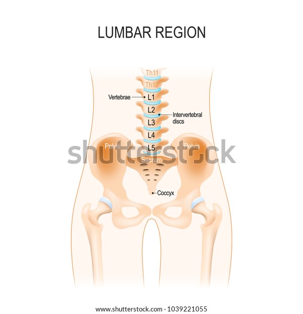 Lumbar Region Anatomy Hip Vertebra Pelvis Stock Vector
Hip pain may result from inflammation degeneration or injury to structures and tissues within the hip joint.

Hip area anatomy. Hip ligaments and tendons tough fibrous tissues that bind bones to bones and muscles to bones. The hip joint is one of the most important joints in the human body. Muscles play an important role in the health and well being.
The hip bones join to the upper part of. Hip in anatomy the joint between the thighbone femur and the pelvis. A healthy hip can support your weight and allow you to move without pain.
Problems within the hip joint itself tend to result in pain on the inside of your hip or your groin. Some of the other muscles in the hip are. It allows us to walk run and jump.
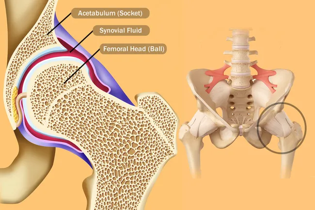
And synovial membrane and fluid which encapsulates the hip joint and lubricates it respectively. Weight bearing stresses on the hip during walking can be 5 times a persons body weight. Hip pain on the outside of your hip upper thigh or outer buttock is usually caused by problems with muscles ligaments tendons and other soft tissues that surround your hip joint.
The hip joint is the uppermost part of the leg where the head of the thigh bone femur fits into the socket of the pelvis. In vertebrate anatomy hip or coxa in medical terminology refers to either an anatomical region or a joint. There are two hip bones one on the left side of the body and the other on the right.
The posterior muscle group is made up of the muscles that extend straighten the thigh at the hip. Amphibians and reptiles have relatively weak pelvic girdles and the femur extends horizontally. The hip joint is a ball and socket joint.
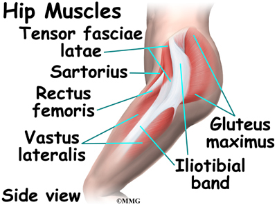
Hip problems occur when any one of these components starts to degenerate or is in some way compromised or irritated. Adductor muscles on the inside of your thigh. Rectus femoris muscle one of the quadriceps muscles on the front of your thigh.
Iliopsoas muscle a hip flexor muscle that attaches to the upper thigh bone. Together they form the part of the pelvis called the pelvic girdle. It bears our bodys weight and the force of the strong muscles of the hip and leg.
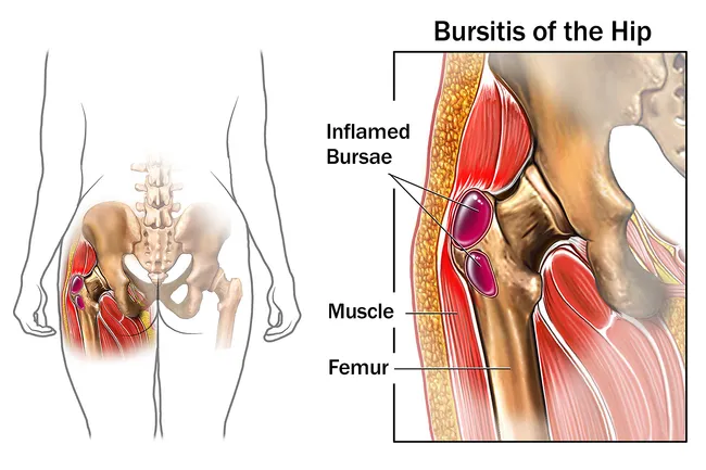
Yet the hip joint is also one of our most flexible joints and allows a greater range of motion than all other joints in the body except for the shoulder. Hip pain may be due to a variety of common causes including fractures sprains strains arthritis and bursitis. Also the area adjacent to this joint.
Hip anatomy function and common problems. The hip joint is one of the largest joints in the body and is a major weight bearing joint. The round head of the femur rests in a cavity the acetabulum that allows free rotation of the limb.
 Hip Anatomy Pictures Function Problems Treatment
 Bones Of The Lower Limb Anatomy And Physiology I
 Hip Anatomy Diagram From Bones To Joints Science Trends
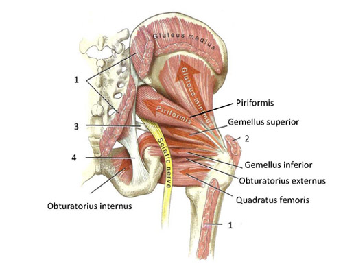 Functional Anatomy Of The Small Pelvic And Hip Muscles
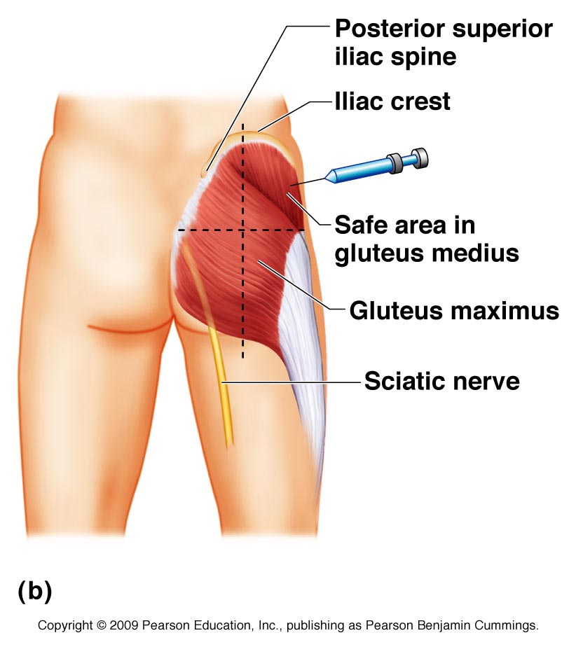 Gluteal Region Anatomy And Significance Bone And Spine
 Hip Flexors An Overview Sciencedirect Topics
:background_color(FFFFFF):format(jpeg)/images/library/11030/Hip_and_thigh_1.png) Hip And Thigh Bones Joints Muscles Kenhub
Hip And Thigh Bones Joints Muscles Kenhub
Hip Labral Tear Cleveland Clinic
 Module 2 Lower Extremity Orthopedic Imaging
 Anatomy And Injuries Of The Hip Anatomical Chart
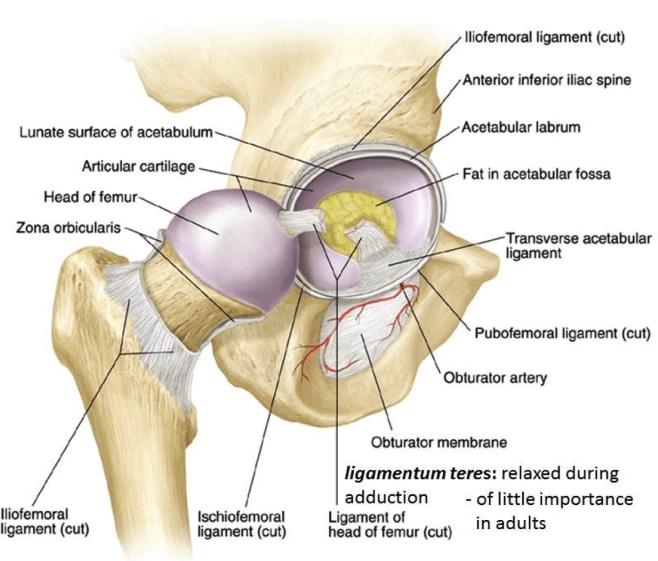 Hip Anatomy Recon Orthobullets
 Chapter 35 Gluteal Region And Hip The Big Picture Gross
 Hip Anatomy Pictures Hip Anatomy Hip Anatomy Muscle
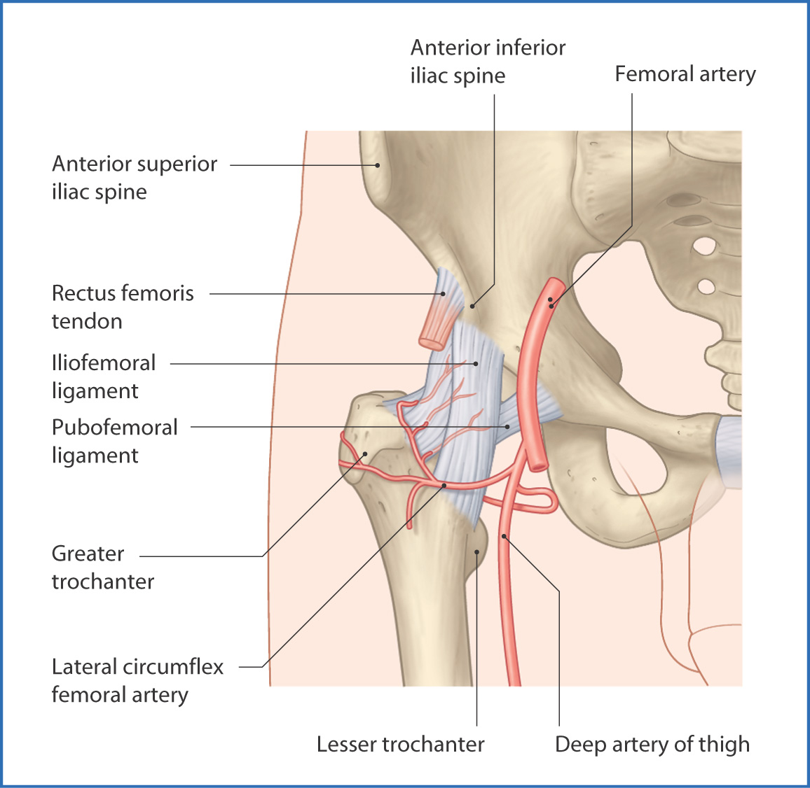 Hip Anatomy Recon Orthobullets
 Muscle Anatomy Of The Hips Buttocks Everything You Need To Know Dr Nabil Ebraheim




Belum ada Komentar untuk "Hip Area Anatomy"
Posting Komentar