Anatomy For Ultrasound
A level 2 ultrasound is a special test that gives you a very specific glimpse of your growing baby. I wasnt ready for how intense my 20 week anatomy scan was.
 On Radiology Normal Anatomy Of Gall Bladder
Some wavelengths are intended to break up tissue but the wavelengths that we use for diagnostics imaging are not at all in the ranges that could cause significant tissue damage.

Anatomy for ultrasound. What is an anatomy ultrasound. For most parents to be the 20 week anatomy ultrasound is their big chance to sneak a peek at their baby before his birth day. These days its pretty much routine for women in their second trimester to be scheduled for a level 2 ultrasound commonly called the 20 week anatomy scan.
Liver ultrasound showing education liver segments normal liver anatomy portal vein hepatic veins the biliary tree and ultrasound scanning protocol worksheets. There are different wavelengths of ultrasounds. When the pregnancy hits the 20th week of gestation an anatomy ultrasound is often ordered.
But this test can do more than let you glimpse your baby. Was that i got a sneak peak at my babies with an ultrasound every time i went to the doctor for a checkup. This sonogram is used to determine fetal anomalies the babys size and weight and also to measure growth to ensure that the fetus is developing properly.
By the end of my pregnancy. Throughout this course the fundamental use and clinical application of ultrasound as a diagnostic tool will be explored through seven key examinations. Other than finding out the sex of your baby if you want to know the ultrasound technician will be.
Color atlas of ultrasound anatomy second edition presents a systematic step by step introduction to normal sectional anatomy of the abdominal and pelvic organs and thyroid gland essential for recognizing the anatomic landmarks and variations seen on ultrasound. The anatomy scan is a level 2 ultrasound which is typically performed between 18 and 22 weeks. The gender of your babybabies can usually be determined at this ultrasound.
Ultrasound means outside of the range of human hearing. In some cases the baby may have their legs crossed or be facing away from the abdomen and thus the sexual organs will not be visible during the anatomic ultrasound. A supplemental anatomy course introducing the diagnostic imaging modality of ultrasound.
 Ultrasound Of The Pancreas What Normal Looks Like
 Scrotal Ultrasound Startradiology
 Ultrasound 3 Anatomy Scan Zygotta
 Ultrasound Of Liver Segments Anatomy
 Ultrasound Examination Of Fetal Anatomy 20 23 Weeks Venus
 Normal Neonatal Head Ultrasound
 Gender Anatomy Development Ultrasound 20 Wks
 Figure 7 From Head And Neck Anatomy And Ultrasound
 Pancreas Anatomy Ultrasound Liver Anatomy Radiology
 Optimizing An Ultrasound Image Hadzic S Peripheral Nerve
 Ultrasound Guided Popliteal Sciatic Block Nysora
 Liver Measurement Ultrasound Pancreas And Its Proportions
 Vikas Shah On Twitter Anatomy Quiz Answer Frcr Foamrad
Anyone Been Told Girl But Really Had A Boy July 2015
 Ultrasound Guided Sciatic Nerve Block Nysora
 Liver Hilum Anatomy Ultrasound

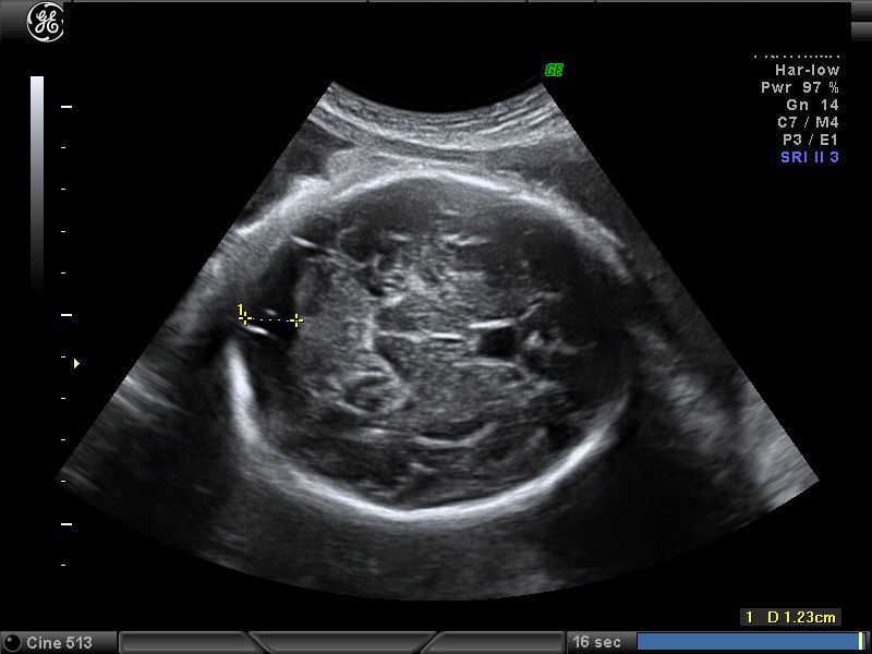 Ultrasound Images Of Fetal Brain
Level 2 Ultrasound 20 Week Anatomy Scan
 Section 7 Atlas Of Ultrasound Guided Anatomy Hadzic S
An Ultrasound For My Birthday Stories Thyme
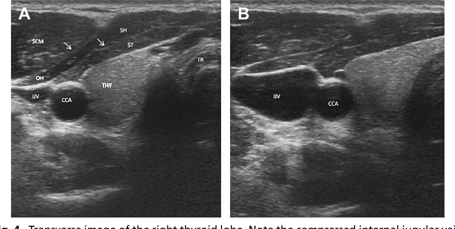 Figure 7 From Head And Neck Anatomy And Ultrasound
 Shoulder Anatomy On Ultrasound Radiology Case
Anatomy Ultrasound Maternal Fetal Associates Of The Mid
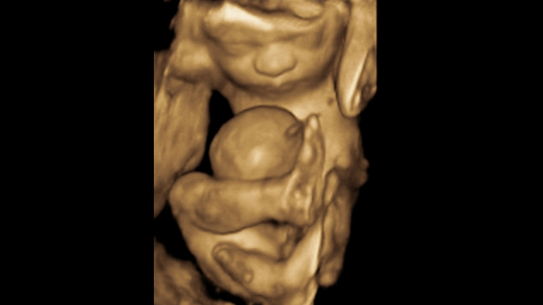 High Resolution Fetal Ultrasound Children S Hospital Of
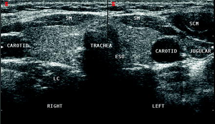 Normal Neck Anatomy And Method Of Performing Ultrasound
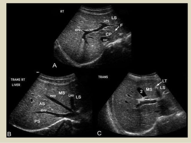 Presentation1 Abdominal Ultrasound Anatomy
 18 20 Week Anatomy Ultrasound Pictures Babycenter
 20 Week Ultrasound Anatomy Scan Lani
 Labelled Fetal Heart Ultrasound
 Cute Clear Ultrasound Of Baby Lucky 19 Weeks Ultrasound Anatomy Scan
 19 Weeks Our Anatomy Ultrasound The Love Notes Blog

Belum ada Komentar untuk "Anatomy For Ultrasound"
Posting Komentar