Anatomy Of Bottom Of Foot
At the same time the foot must be strong to support more. Dorsal refers to the top surface of the foot whereas plantar takes its name from the fact that the foot is planted on the ground when it is in contact with a surface.
 Ankle Block Landmarks And Nerve Stimulator Technique Nysora
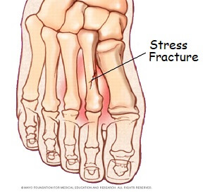
The metatarsals which run through the flat part of your foot.

Anatomy of bottom of foot. Dorsal and plantar see fig. The feet are divided into three sections. These arches the medial arch lateral arch and fundamental longitudinal arch are created by the angles of the bones and strengthened by the tendons.
Therefore plantar refers to the bottom aspect of the foot. The hindfoot forms the heel and ankle. The foot is an extremely complex anatomic structure made up of 26 bones and 33 joints that must work together with 19 muscles and 107 ligaments to execute highly precise movements.
The anatomy of the foot anatomi cally the foot has two surfaces. Below the juncture of these bones are the arches of the foot which are three curves at the bottom of the foot that makes walking easier and less taxing for the body. The talus which is the.
Calcaneus the largest bone of the foot which lies beneath the talus to form the heel bone. The midfoot is a pyramid like collection of bones that form the arches of the feet. The forefoot contains the five toes phalanges and the five longer bones metatarsals.
Tarsals five irregularly shaped bones of the midfoot that form the foots arch. Talus the bone on top of the foot that forms a joint with the two bones of the lower leg. The cuneiform bones the navicularis and the cuboid all of which function to give your foot.
The end of the leg on which a person normally stands and walks. The talus bone supports the leg bones. The calcaneus which is the bone in your heel.
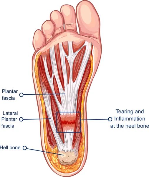
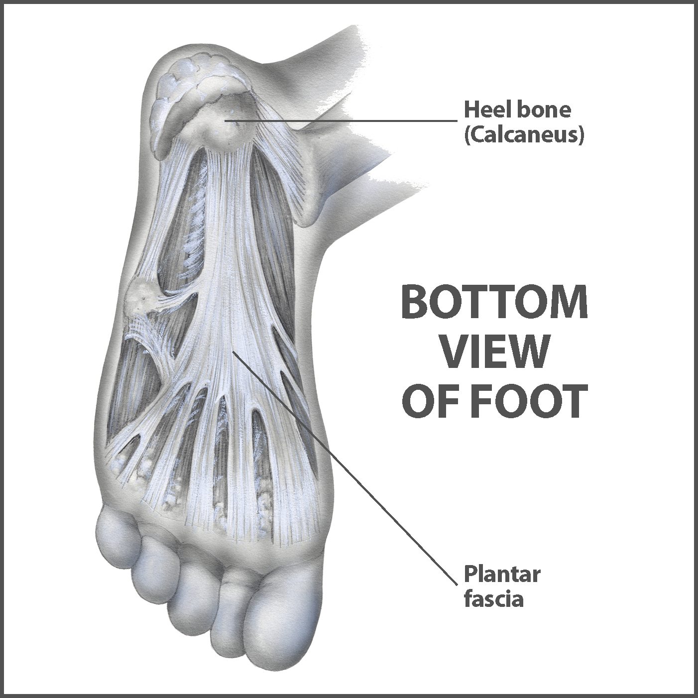
The phalanges which are the bones in your toes. For instance the plantar fascia is an important structure on the bottom of the foot that is important for both the normal function of the foot and for maintaining the normal arch of the foot. The bones of the feet are.
Picture of foot anatomy detail. It is also a common cause of heel pain also known as plantar fasciitis.
 ᐈ The Bottom Of Your Foot Stock Pictures Royalty Free
:max_bytes(150000):strip_icc()/heelpainfinal-01-5c86a48246e0fb00014319ff.png) Heel Pain Causes Treatment And When To See A Doctor
Heel Pain Causes Treatment And When To See A Doctor
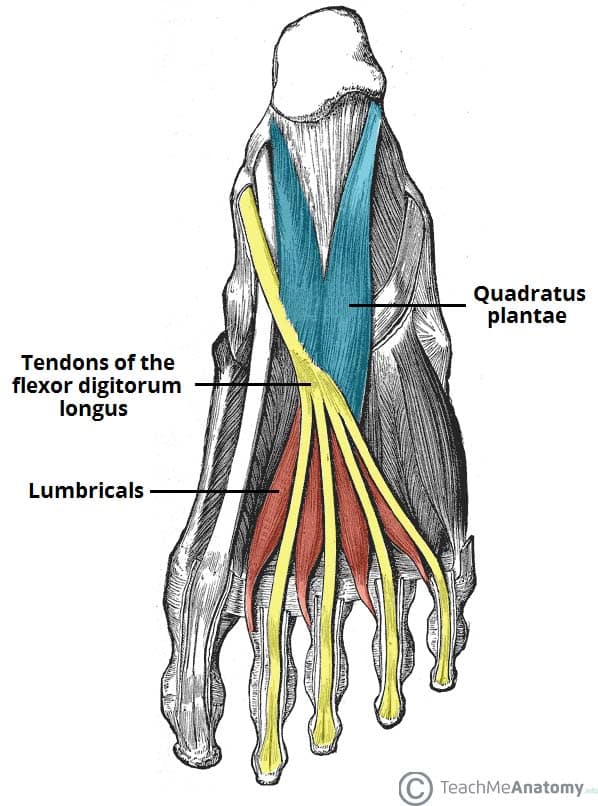 Muscles Of The Foot Dorsal Plantar Teachmeanatomy
 Nerves Of The Leg Foot Everything You Need To Know Dr Nabil Ebraheim
 Anatomical Overlays Bottom Of The Foot These Images Will
 The Newborn Foot American Family Physician

 Anatomy Bottom View Foot Stock Illustrations 44 Anatomy
 Foot Anatomy Bones Ligaments Muscles Tendons Arches
 Anatomy Of The Foot Footmaxx
 Foot Anatomy Bones Bottom View Fa12 Future Nurse Foot
 Ball Of Foot Pain Do The Bottoms Of Your Feet Toes Hurt

 Tight Shoes Commonly Associated Foot Conditions Kintec
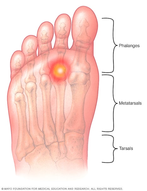 Metatarsalgia Symptoms And Causes Mayo Clinic
 How To Release Your Plantar Fascia Helps Plantar Fasciitis
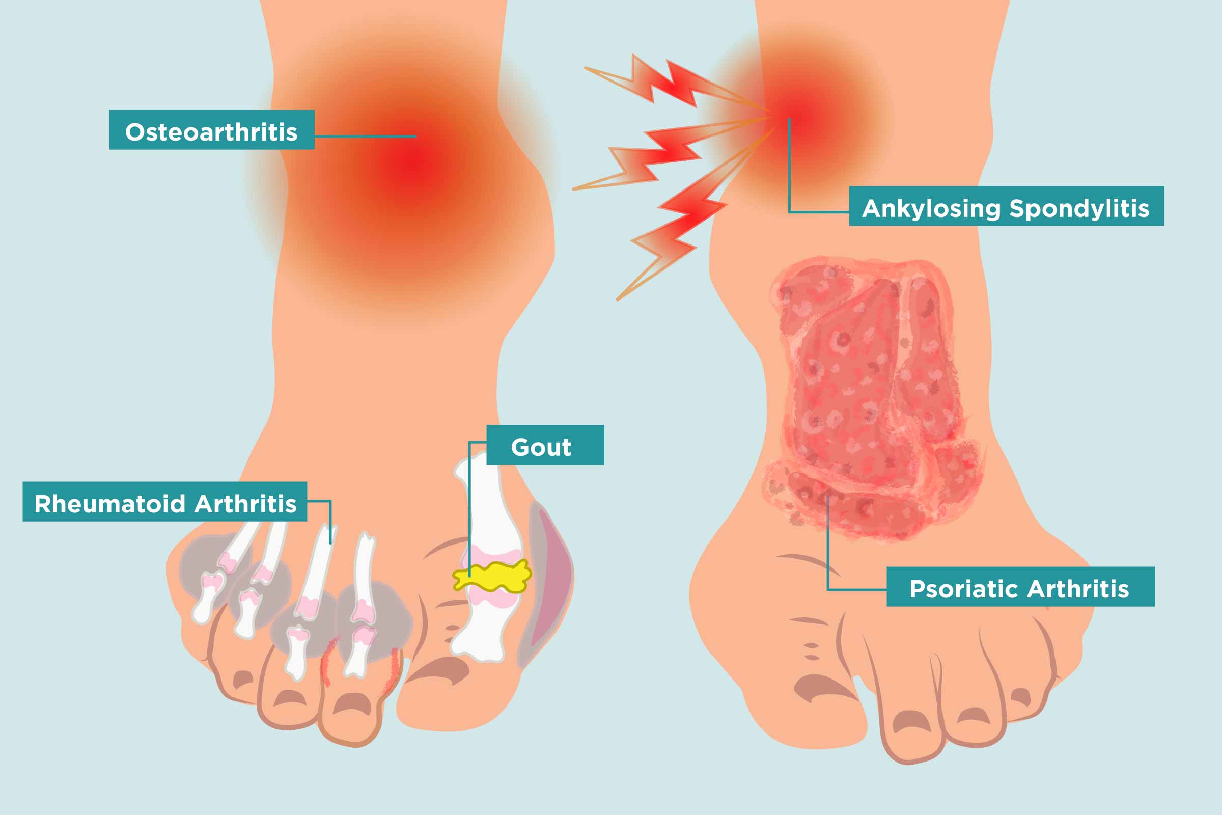 Arthritis In Your Feet Causes Symptoms And Treatment
 Ball Of Foot Pain Do The Bottoms Of Your Feet Toes Hurt
 Ball Of Foot Pain Relief Management Dr Scholl S

 Sole Foot Wikipedia
 Foot Pain Identifier Foot Com
 Foot Arch Pain Causes Treatment Foot Pain Explored
 Layers Of The Plantar Foot Foot Ankle Orthobullets
 Foot Pain Diagnosis Achilles Tendinitis Causes Home
 Plantar Fasciitis Info Florida Orthopaedic Institute
 Understanding Heel Pain
 Anatomy 101 Strengthen Your Big Toes To Build Stability
 Foot Pain Diagnosis Achilles Tendinitis Causes Home
 Plantar Fasciitis Treatment Relief For Plantar Fasciitis
 Foot Anatomy Images Stock Photos Vectors Shutterstock
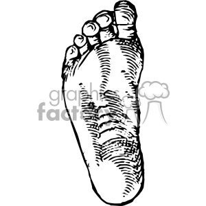 Cousin Jehan Vintage Bottom Of Foot Vector Anatomy Art Clipart Royalty Free Clipart 403137
 Nerves Of The Leg And Foot Interactive Anatomy Guide
Belum ada Komentar untuk "Anatomy Of Bottom Of Foot"
Posting Komentar