Anatomy Heart Diagram
It is the simple squamous endothelium layer which lines the inside of the. The heart is the epicenter of the circulatory system which supplies the body with oxygen and other important nutrients needed to sustain life.
 Anatomy Of The Human Heart Posterior View Purposegames
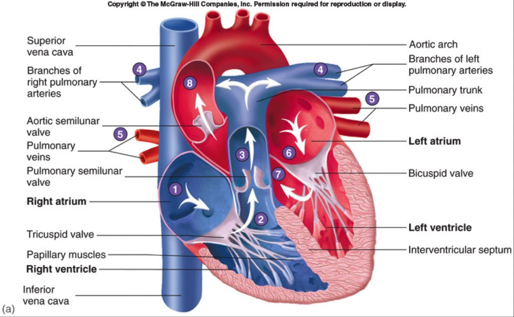
The right atrium receives blood from the veins and pumps it to the right ventricle.
Anatomy heart diagram. Without the heart the tissues couldnt get the oxygen they need and would die. Because the heart points to the left about 23 of the hearts mass is found on the left side of the body and the other 13 is on the right. Anatomy of the heart pericardium.
This amazing muscle produces electrical impulses that cause the heart to contract. The heart pumps blood through the network of arteries and veins called the cardiovascular system. The heart has four chambers.
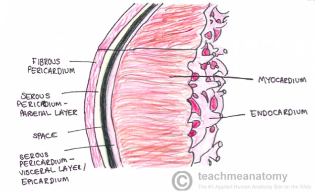
A double layered membrane called the pericardium surrounds your heart like a sac. Your heart is located between your lungs in the middle of your chest behind and slightly to the left of your breastbone sternum. Along with lymphatic vessels the blood blood vessels and lymph the heart composes the circulatory system of the body.
Lets examine the anatomy of the heart along with some diagrams that show how the heart operates. Structure of heart wall in human heart diagram epicardium. The right ventricle receives blood from the right atrium and pumps it to the lungs where it is loaded with oxygen.
The heart is situated within the chest cavity and surrounded by a fluid filled sac called the pericardium. The heart sits within a fluid filled cavity called the pericardial cavity. The heart is a mostly hollow muscular organ composed of cardiac muscles and connective tissue that acts as a pump to distribute blood throughout the bodys tissues.
The walls and lining of the pericardial cavity are a special membrane known as the pericardium. The heart weighs between 7 and 15 ounces 200 to 425 grams and is a little larger than the size of your fist. The myocardium is muscular middle layer of heart wall which contain.
The epicardium is one of the most outer layers of the heart wall. It is divided by a partition or septum into two halves and the halves are in turn divided into four chambers. The anatomy of the heart.
:max_bytes(150000):strip_icc()/human-heart-circulatory-system-598167278-5c48d4d2c9e77c0001a577d4.jpg) The Anatomy Of The Heart Its Structures And Functions
The Anatomy Of The Heart Its Structures And Functions
 Human Heart Gross Structure And Anatomy Online Biology Notes
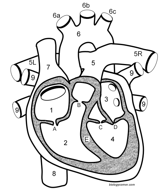
 Anatomy Of A Human Heart
 Heart Valves Yourheartvalve
 Body Scientific International Post It Anatomy Of Heart Chart Teaching Supplies Classroom Safety
 The Heart Teachmeanatomy
 1 Heart Anatomy From The Anterior View Left And Interior
 Anatomy Heart External
 Human Heart Diagram Labeled Science Trends
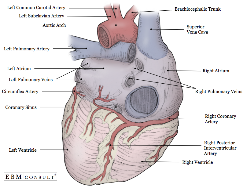
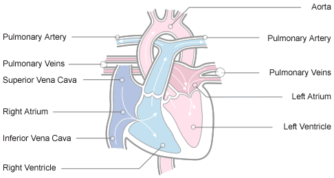
 Anatomy And Function Of The Coronary Arteries Johns
Anatomy And Function Of The Heart S Electrical System
 Learn The Anatomy Of The Heart
 Anatomy And Physiology Of The Heart Normal Function Of The
 External Anatomy Of Heart Heart Anatomy Heart Valves
 Human Heart Anatomy Diagram Function Chambers Location
 Anatomical Diagrams Of Heart Heart Failure Online
 6 The Heart
![]() Heart Anatomy Structure Valves Coronary Vessels Kenhub
Heart Anatomy Structure Valves Coronary Vessels Kenhub
 Jolie Blogs Heart Diagram Anatomy
 Heart Wikipedia
Human Heart Diagram Photos 11 578 Human Heart Stock Image
 1 Shows The Heart Anatomy From The Anterior And Interior
![]() Anatomy Of The Human Heart Teachervision
Anatomy Of The Human Heart Teachervision
 Free Art Print Of Human Heart Anatomy
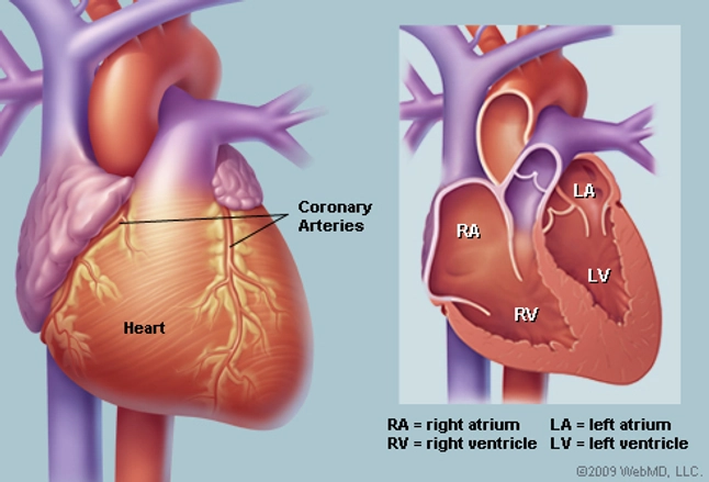
 Heart Valve Anatomy Britannica
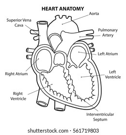
 Heart Diagram Right Left Atria Right Left Ventricles
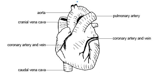 Anatomy And Physiology Of Animals Cardiovascular System The
:max_bytes(150000):strip_icc()/heart_exterior_anatomy-577d5cc23df78cb62c942f06.jpg) The Anatomy Of The Heart Its Structures And Functions
The Anatomy Of The Heart Its Structures And Functions
 Anatomy Of The Heart Emt Training Station
 Anatomy Of The Human Heart

Belum ada Komentar untuk "Anatomy Heart Diagram"
Posting Komentar