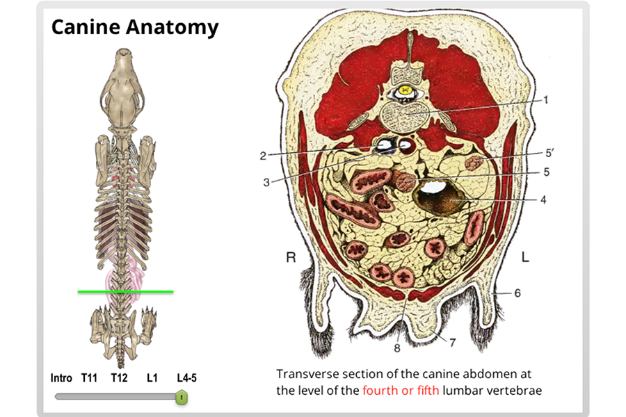
Canine Abdomen Anatomy
The urinary system is responsible for removing waste products from blood and eliminating them as urine. Only the tissues size does.
 Dog Muscular Skeletal Nerves Canine Anatomy Poster 24 X 36 Veterinary Chart
The esophagus can therefore be referred to as having cervical thoracic and abdominal portions.
Canine abdomen anatomy. Hematopoiesis organ that produces lymphocytes. The flank refers to the side of the dog between the end of the chest and the rear leg. If the dog is thin or deep chested the caudal abdominal horizontal line will be higher more dorsal than after taking the cranial abdominal lateral view.
Seat of the intelluctual capacities of a gog. Virtual canine anatomy is an innovative anatomy program that has received outstanding accolades from members of the american association of veterinary anatomists students and instructors both in the united states and internationally. Important part of the nervous system.
This veterinary anatomical atlas includes selected labeling structures to help student to understand and discover animal anatomy skeleton bones muscles joints viscera respiratory system cardiovascular system. As they grow bigger in size the number of these bones muscles or tendons does not increase. The genital organs are involved in reproduction.
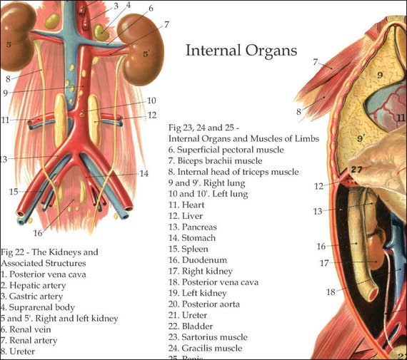
The urogenital system cat dog includes the kidneys ureters urinary bladder urethra and the genital organs of box sexes. Anatomy of the male canine abdomen and pelvis on ct imaging this module of the vet anatomy veterinary atlas concerns the abdomen and pelvis of the dog in ct. A muscular tube that carries ingesta from the laryngopharynx to the stomach.
Anatomy of the dog illustrated atlas this modules of vet anatomy provides a basic foundation in animal anatomy for students of veterinary medicine. Internal anatomy of a dog. The anatomy of the canine digestive system an overview esophagus.
Ct images are from a healthy 6 year old castrated male dog. A dogs anatomy is not very different from any other mammals. When the pups are born they have all the bones muscles and tendons that an adult dog has.
Remember to always include the sternum and xiphoid process in order to include cranial abdominal anatomy. Carnivorous domestic mammal raised to perform various tasks for humans. The loin is the back between the end of the rib cage and the beginning of the pelvic bone.
The esophagus courses through the neck thorax and into the abdomen. Baring the canine back and chest. The belly or abdomen is the underside of the dog from the end of its rib cage to its tail.
Part of the digestive tract between the esophagus and the intestine.
 Abdomen And Pelvis Anatomy Of The Dog On Ct
 2019 Ultimate Veterinary Guide To Dog Anatomy With Images
 Canine Abdominal Organs Ventral View Unlabeled Clipart
 Canine Anatomy Design Group Vet Med
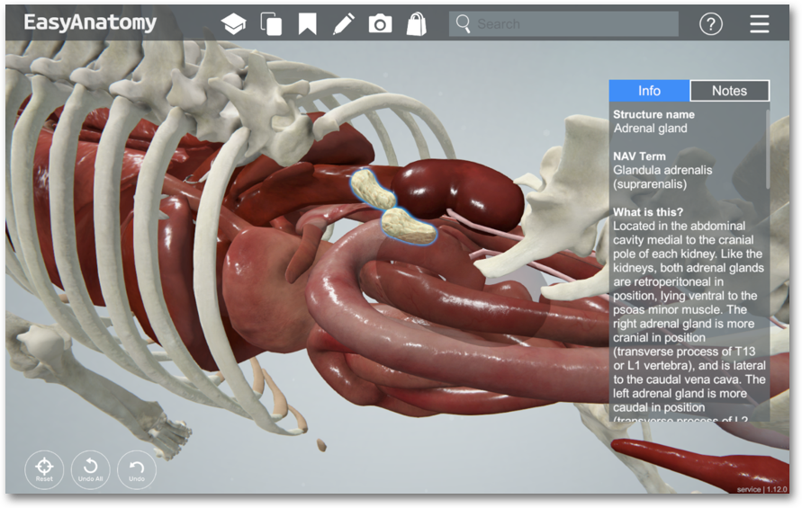
What Is Addison S Disease In Dogs Easyanatomy
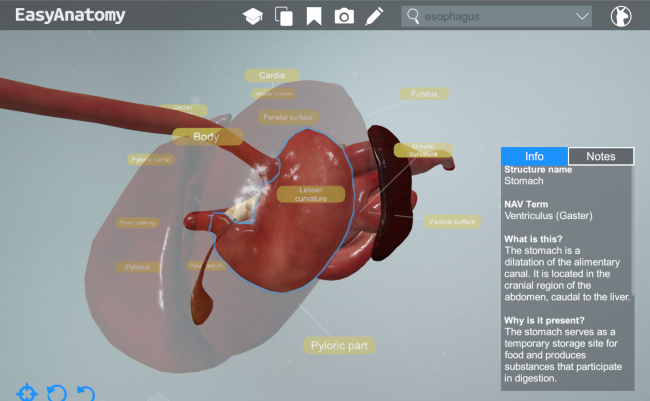
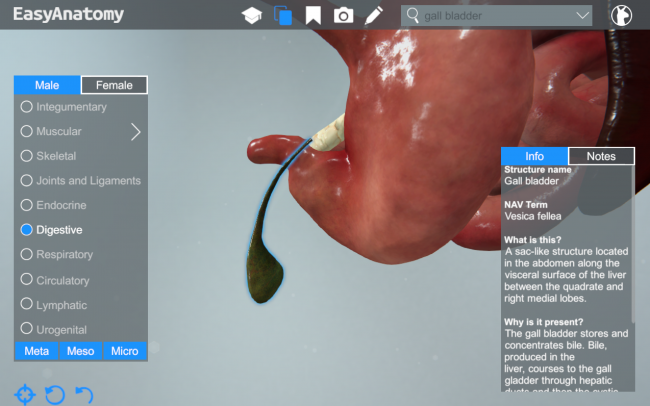 Anatomy Of The Canine Digestive System Easyanatomy
 Abdominal Cavity Anatomy Britannica
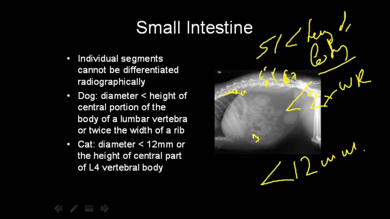 Vet Talks Normal Radiographic Anatomy Of The Canine Abdomen
 Ventral View Of The Abdomen Of The Dog Showed The
 Esophagus Veterian Key
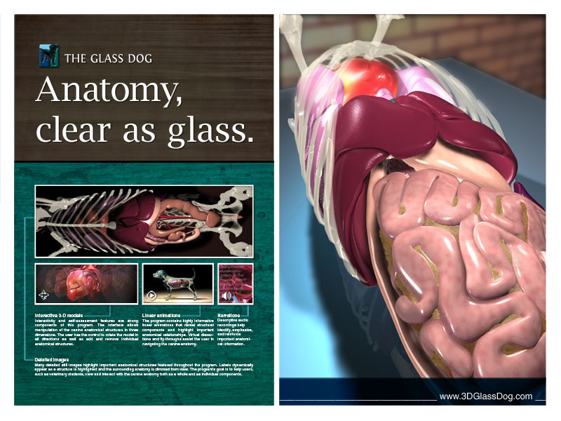 Gilleland Medical Illustration The Glass Dog
 Surgical Views Suspensory Ligament Rupture Technique
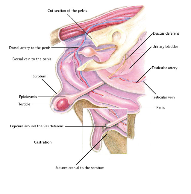
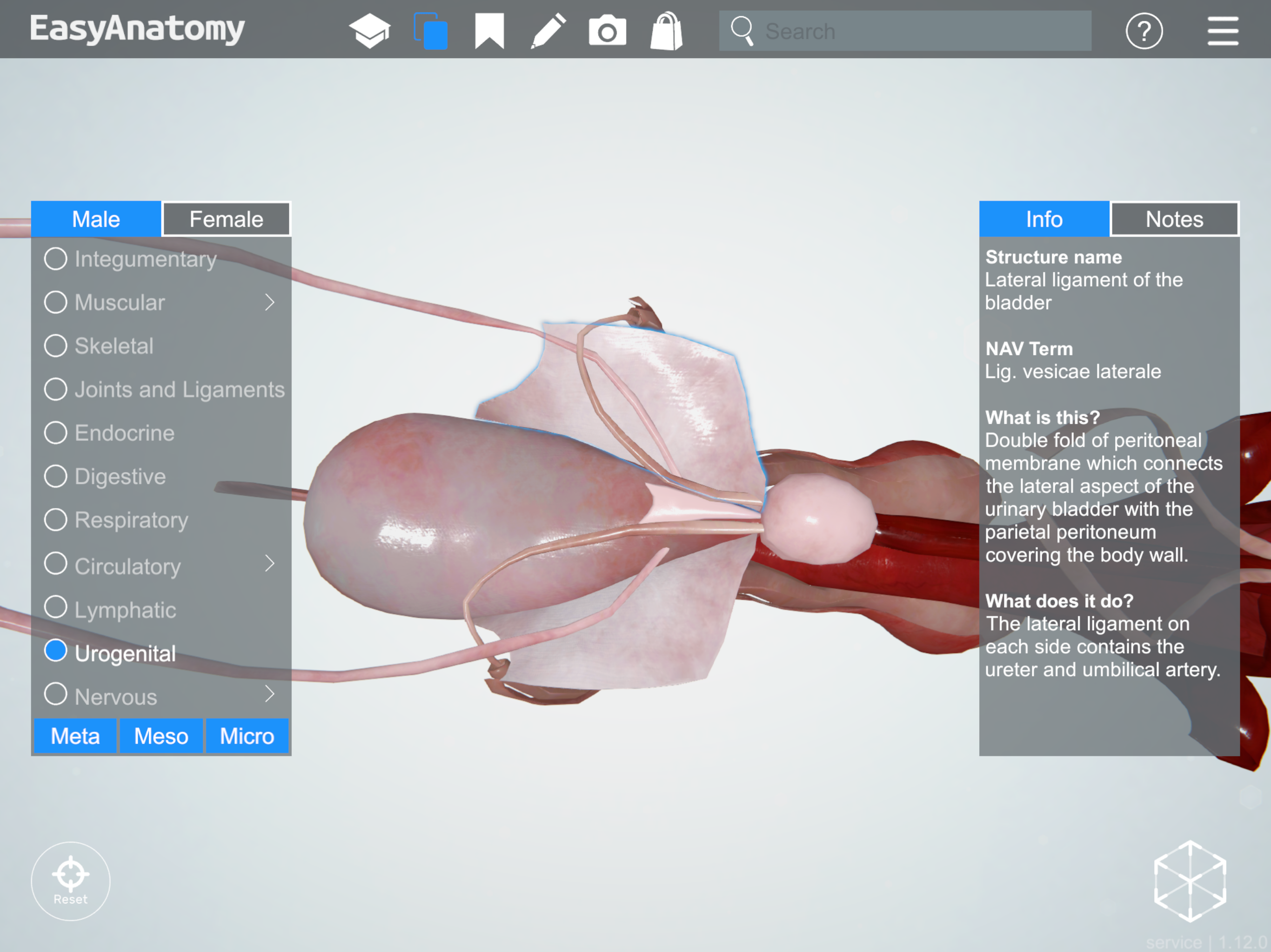 Anatomy Of The Canine Male Urogenital System Easyanatomy
 Abdomen And Pelvis Anatomy Of The Dog On Ct
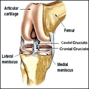 Cranial Cruciate Ligament Injury Metropolitan Veterinary
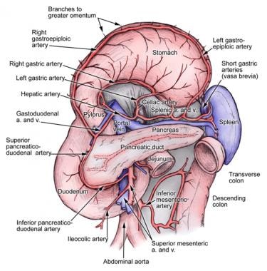 Spleen Anatomy Overview Gross Anatomy Microscopic Anatomy
 Spleen Anatomy Physiology Wikivet English
 Stomach Veterian Key
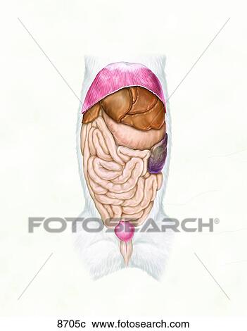
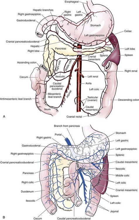
 Dog Anatomy Thoracic And Abdominal Organs
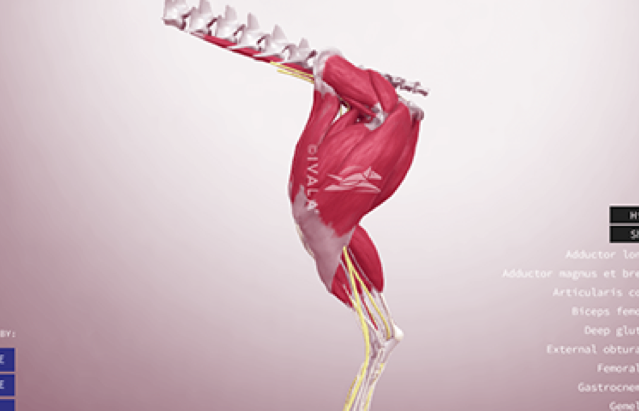 Ivala Learn 3d Veterinary Anatomy Clinical Learning Content
 Anatomy Of The Canine Digestive System Easyanatomy
 Thorax Of The Dog Cross Sectional Anatomy On Computed
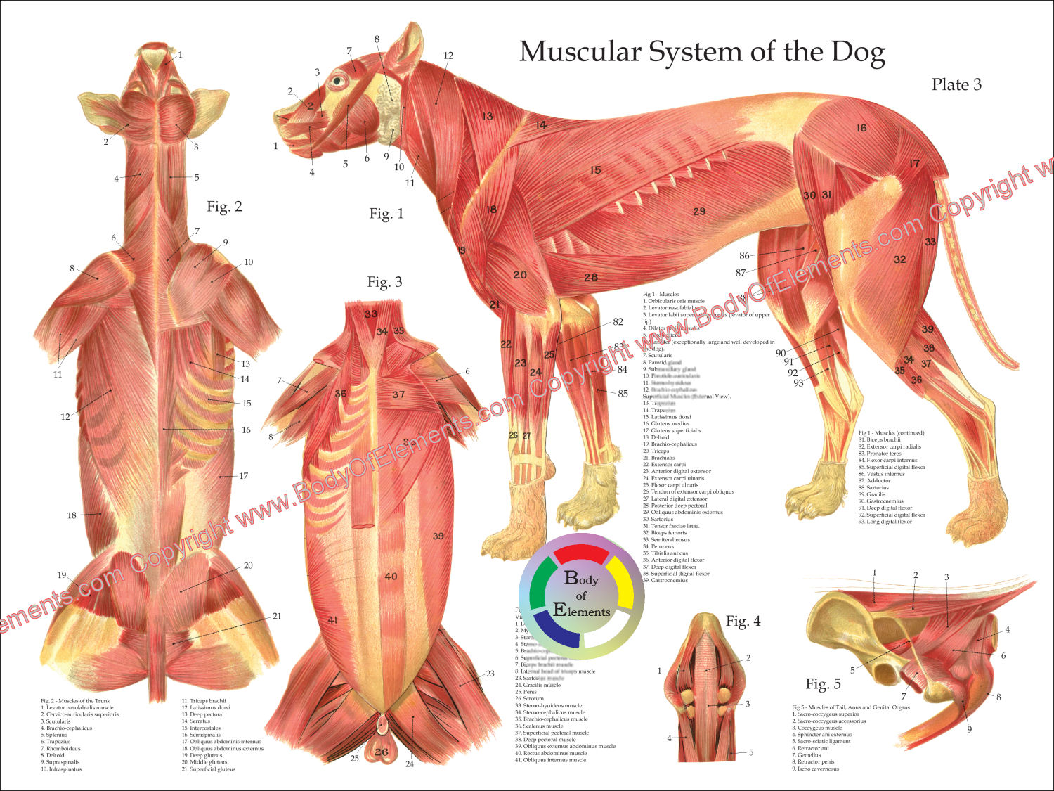 Dog Muscular Anatomy Poster 18 X 24
 Surface Anatomy Wikipedia


Belum ada Komentar untuk "Canine Abdomen Anatomy"
Posting Komentar