Wrist Xray Anatomy
Atlas of wrist mri anatomy. Imaging key wrist ligaments.
There are numerous ligaments but included below are the most clinically significant.

Wrist xray anatomy. Oval surface of one bone fits into an elliptical cavity of another. Trauma x ray upper limb wrist x ray scaphoid fractures. Unable to process the form.
The intrinsic and extrinsic wrist ligaments play a vital role in the stability of the wrist joint. The scaphoid bone is the most commonly fractured wrist bone. Wrist ligaments are best assessed with dedicated wrist mri.
Radiographic anatomy of the skeleton. 14 flexor carpi radialis tendon. Bartolotta 2 michael l.
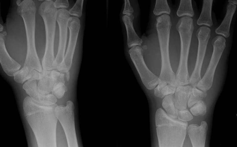
Normal wrist x ray in an adult female for reference. Check for errors and try again. 6 extensor pollicis longus tendon.
90to the fingers and is of particular importance to the dexterity of the hand. 9 extensor carpi radialis longus t. Wrist radiograph an approach dr yair glick and dr jeremy jones et al.
Allan 4 audio available share claim cmesam. Intrinsic ligaments only attach to carpal bones scapholunate ligament. 4 extensor digiti minimi t.
Bateni 1 roger j. Use the mouse to scroll or the arrows. If a carpal bone injury is suspected and not visible on the pa or lateral image.
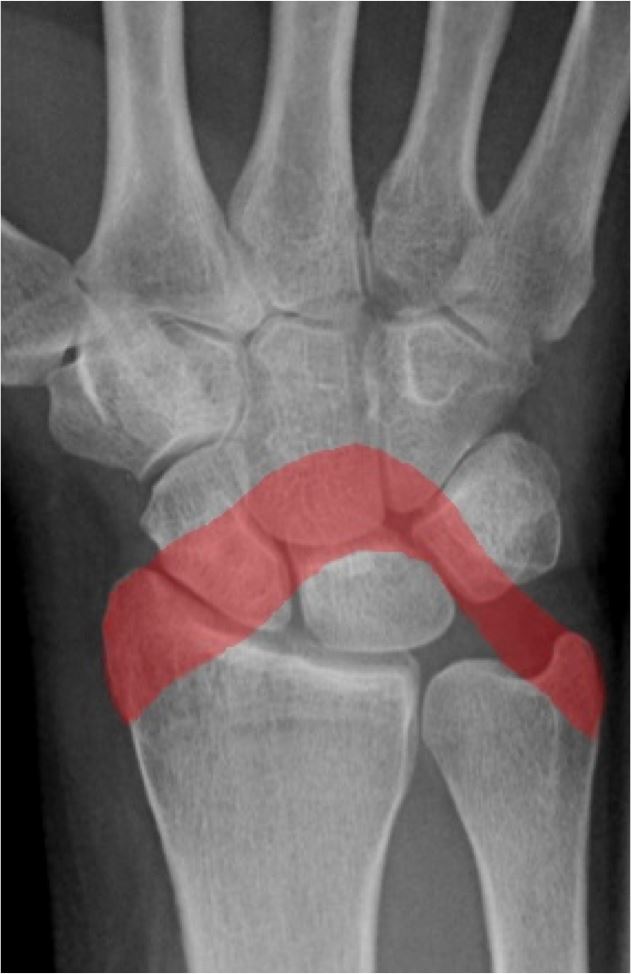
Wrist radiographs are ubiquitous on any night of the week in emergency departments especially when pavements are icy. Functional position of the wrist and hand has been determined to be. Normal radiographic anatomy of the wrist.
What the surgeon needs the radiologist to know cyrus p. 3 extensor carpi ulnaris t. There are numerous joints of the wrist named according to their.
Knee shoulder shoulder arthrogram ankle elbow wrist hip contact. 8 extensor carpi radialis brevis t. Biaxial typically flexionextension and abductionadduction.
5 extensor digitorum indicis tt. Richardson 3 hyojeong mulcahy 3 and christopher h. Copyright c 2005 2019 alex freitas md.
 Hd Radiology Review Hand And Wrist X Rays Em Rap
 Wrist Dislocation An Overview Sciencedirect Topics

 Hand X Ray Medical Art Library
Radiology Anatomy Images Wrist X Ray Radiographic Anatomy
 Colles Fracture Lateral A And Ap B Wrist Radiographs
 How To Take And Interpret Avian Radiographs Veterinary

 Film Critique Of The Upper Extremity Part 3 Hand Wrist
 The Radiology Assistant Wrist Carpal Instability
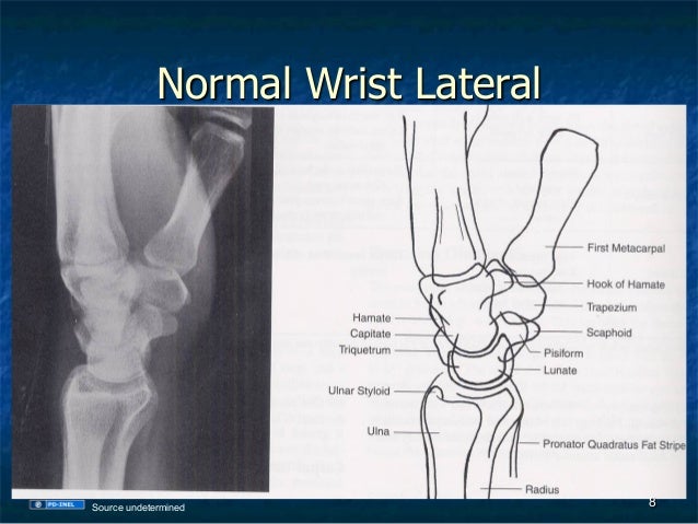 Gemc Radiology X Rays Of The Hand And Wrist Resident Training
 Radiological Anatomy Of The Shoulder Arm Elbow Forearm
A Radiologist S Guide To Wrist Alignment
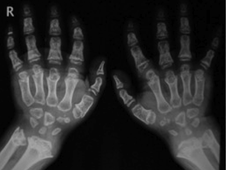 Bone Of Contention Can Wrist X Rays Really Reveal The Age
Bone Age A Handy Tool For Pediatric Providers
 A Normal Wrist Xray Geriatric Occupational Therapy
 Steph Curry Gets Surgery For Broken Left Hand This Is What
 Plain Wrist Radiographs Of The Patient On The Symptomatic
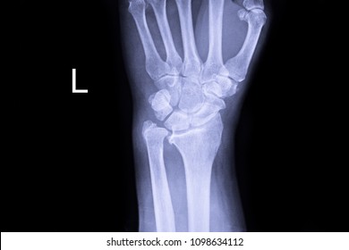 Wrist Xray Images Stock Photos Vectors Shutterstock
 Distal Radius Fracture Wikipedia
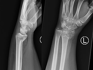


Belum ada Komentar untuk "Wrist Xray Anatomy"
Posting Komentar