Anatomy Of Knee Muscles
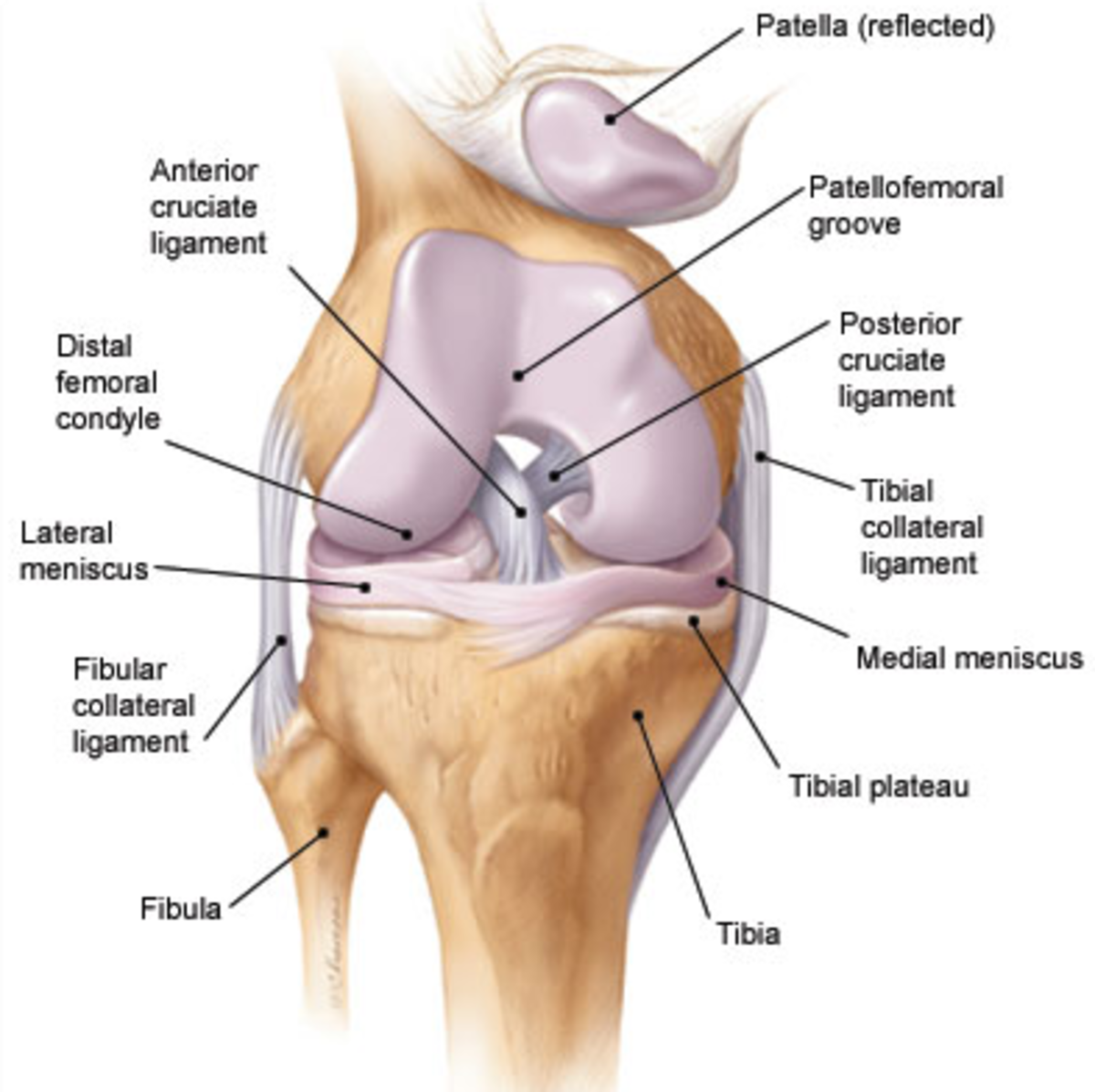
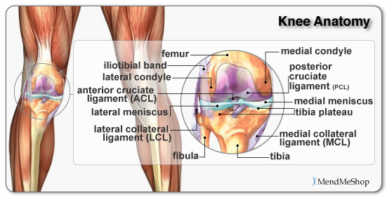
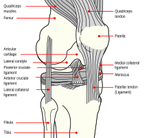
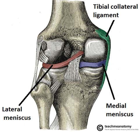
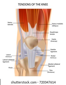
It is formed by articulations between the patella femur and tibia. These are crescent shaped discs that act as a cushion.
 Knee Joint Anatomy Bones Ligaments Muscles Tendons Function
These muscles work in groups to flex extend and stabilize the knee joint.
/188058334-crop-56aae7425f9b58b7d0091480.jpg)
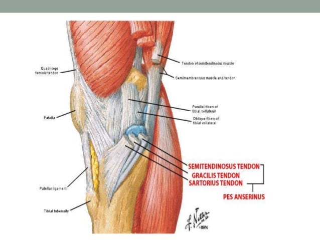
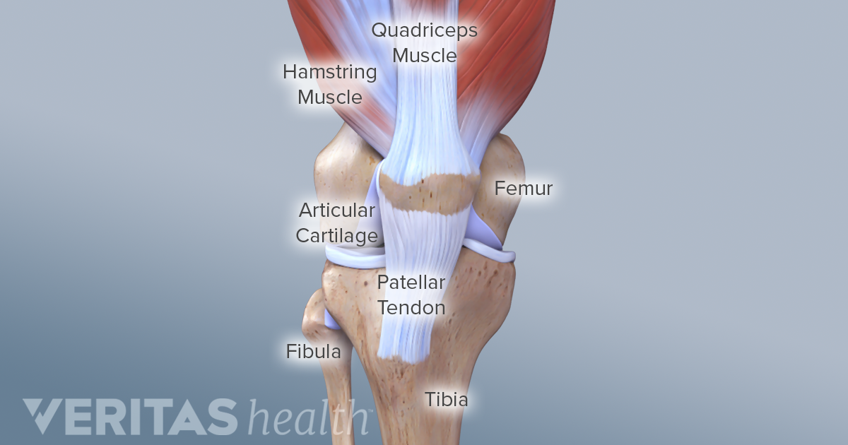
Anatomy of knee muscles. This long muscle extends from the pelvis to the tibia. Ligaments are tough and fibrous tissues. Knee joint anatomy involves looking at each of the different structures in and around the knee.
Each muscle as an origin and insertion. Ligaments are structures that connect two bones together. Tendons connect the knee bones to the leg muscles that move the knee joint.
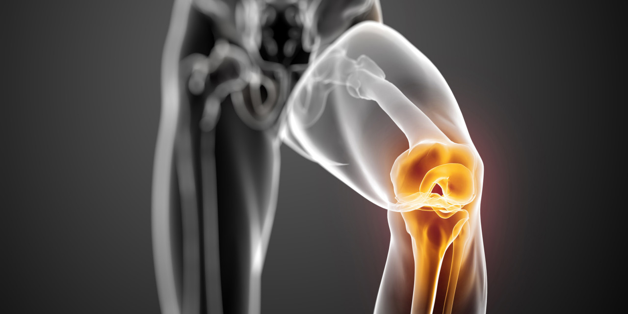
Cartilage of the knee. The knee joins the thigh bone femur to the shin bone tibia. 1 the tibiofemoral joint where the tibia meet the femur 2 the patellofemoral joint where the kneecap or patella meets the femur.
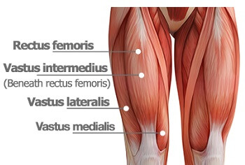
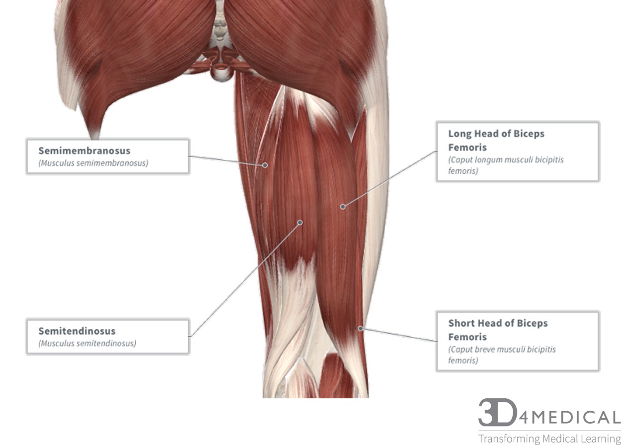
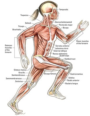
The muscles of the knee include the quadriceps hamstrings and the muscles of the calf. It begins in the thigh area and extends to the head. This muscle also extends the thigh and flexes the knee but the tendons connecting it to.
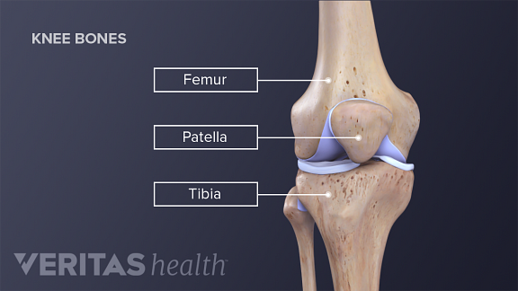
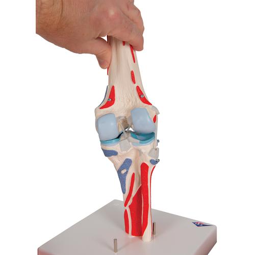
The knee joint is the largest and one of the most complex joints in the human body. They act like strong ropes to connect bones. The femur thigh bone tibia shin bone and patella.
These tough bands of soft tissue. There are three bones that come together at the knee joint. Ligaments of the knee.
4 medial head gastrocnemius muscle. The knee joint is a synovial joint which connects the femur thigh bone the longest bone in the body to the tibia shin bone. T2 weighted fatsat axial view.
There are various muscles that control movement ligaments that give stability special cartilage to absorb pressure and various other structures to ensure smooth pain free movement. There are two types of cartilage of the knee joint. Mri of the knee.
The knee is one of the largest and most complex joints in the body. This long muscle flexes the knee. Atlas of knee mri anatomy.
These motions of the knee allow the body to perform such important movements as walking running kicking and jumping. 6 medial head gastrocnemius muscle. The smaller bone that runs alongside the tibia fibula and the kneecap patella are the other bones that make the knee joint.
There are two main joints in the knee. Knee muscles consist of the quadriceps at the front of the thigh and the hamstring muscles at the back. 7 plantar and lateral head gastrocnemius muscle.
Anatomy of the knee bones around the knee. The knee joint is a hinge type synovial joint which mainly allows for flexion and extension and a small degree of medial and lateral rotation.
 Preventing Acl Injury Through Strengthening Exercises
 Knee Exercises For Arthritis
 Knee Muscles Knee Pain Explained
 Medial Patellofemoral Ligament Mpfl Reconstruction
 Knee Injuries For Parents Nemours Kidshealth
 Knee Anatomy
 Anatomy Of The Knee Bones Muscles Arteries Veins Nerves
Common Knee Injuries Orthoinfo Aaos
 The Mighty Hamstring Muscles Anatomy Injury Training
 The Anatomy Of The Knee Health Fitness Anatomy Of The
What Is Causing Your Knee Pain
 Human Knee Joint Model With Removable Muscles 12 Part 3b Smart Anatomy
 Knee Joint Anatomy Bones Cartilages Muscles Ligaments
 Gastrocnemius Muscle Anatomy Britannica
 Anatomy And Examination Of The Knee
 Knee Leg Atlas Of Anatomy
 Removing Part Of The Meniscus Increases Unwanted Muscle
 Knee Wikipedia
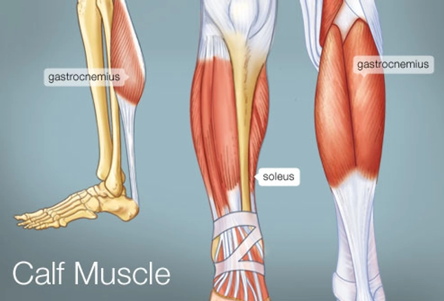 The Calf Muscle Human Anatomy Diagram Function Location
 Knee Joint Picture Image On Medicinenet Com
 Muscles Advanced Anatomy 2nd Ed
 Anatomy Flashcards Knee And Leg Learn All Bones Ligaments
Collateral Ligament Injuries Orthoinfo Aaos
 The Knee Joint Articulations Movements Injuries
 Muscles That Move The Knee And Ankle Dummies
 Imagenes Fotos De Stock Y Vectores Sobre Knee Muscles
 Knee Anatomy
Belum ada Komentar untuk "Anatomy Of Knee Muscles"
Posting Komentar