Anatomy Of The Foot Arch
The foot has three arches. In h the foot is a part of vertebrate anatomy which serves the purpose of supporting the animals weight and allowing for locomotion on land.
 Metatarsalgia Happy Feet Plus Footwear For A Healthier You
The main muscles of the foot are.
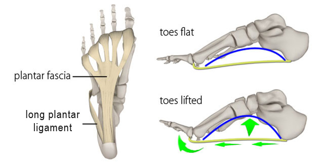
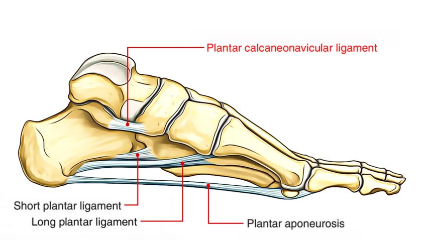
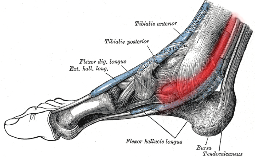
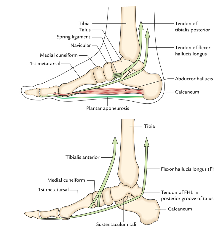
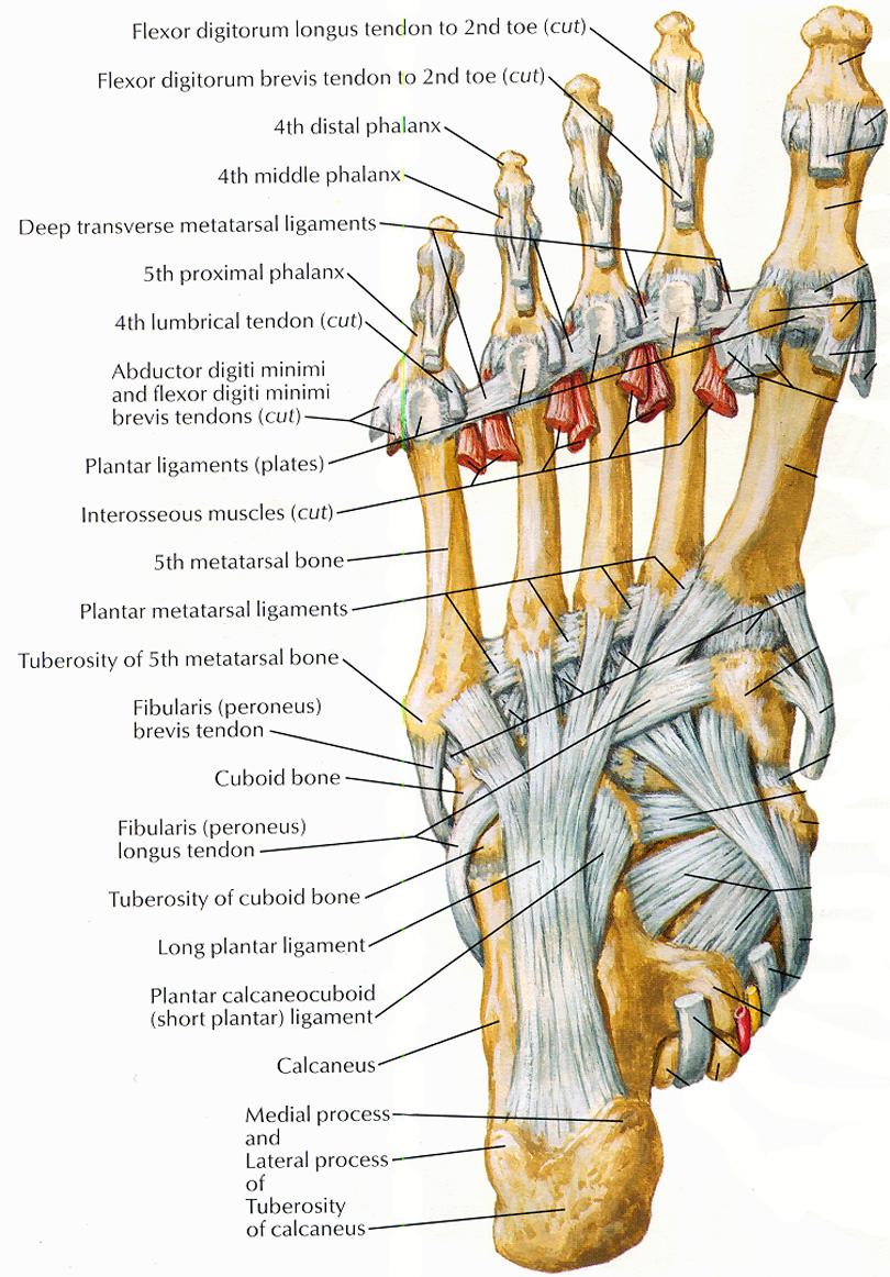
Anatomy of the foot arch. The arch is further supported by the plantar aponeurosis by the small muscles in the sole of the foot by the tendons of the tibialis anterior and posterior and peronæus longus and by the ligaments of all the articulations involved. They are formed by the tarsal and metatarsal bones and supported by ligaments and tendons in the foot. This arch created by the metatarsals is not as obvious as the large medial arch but is still fundamental for distributing weight as we push off from the ground when walking or running.
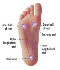
These arches the medial arch lateral arch and fundamental longitudinal arch are created by the angles of the bones and strengthened by the tendons that connect the muscles and the ligaments that connect the bones. Two longitudinal medial and lateral arches and one anterior transverse arch. These include the three cuneiform bones the cuboid bone and the navicular bone.
Arches of the foot the foot is the region of the body distal to the leg and consists of 28 bones. The tibilias peroneal which controls movement on the outside of the ankle. The midfoot is a pyramid like collection of bones that form the arches of the feet.
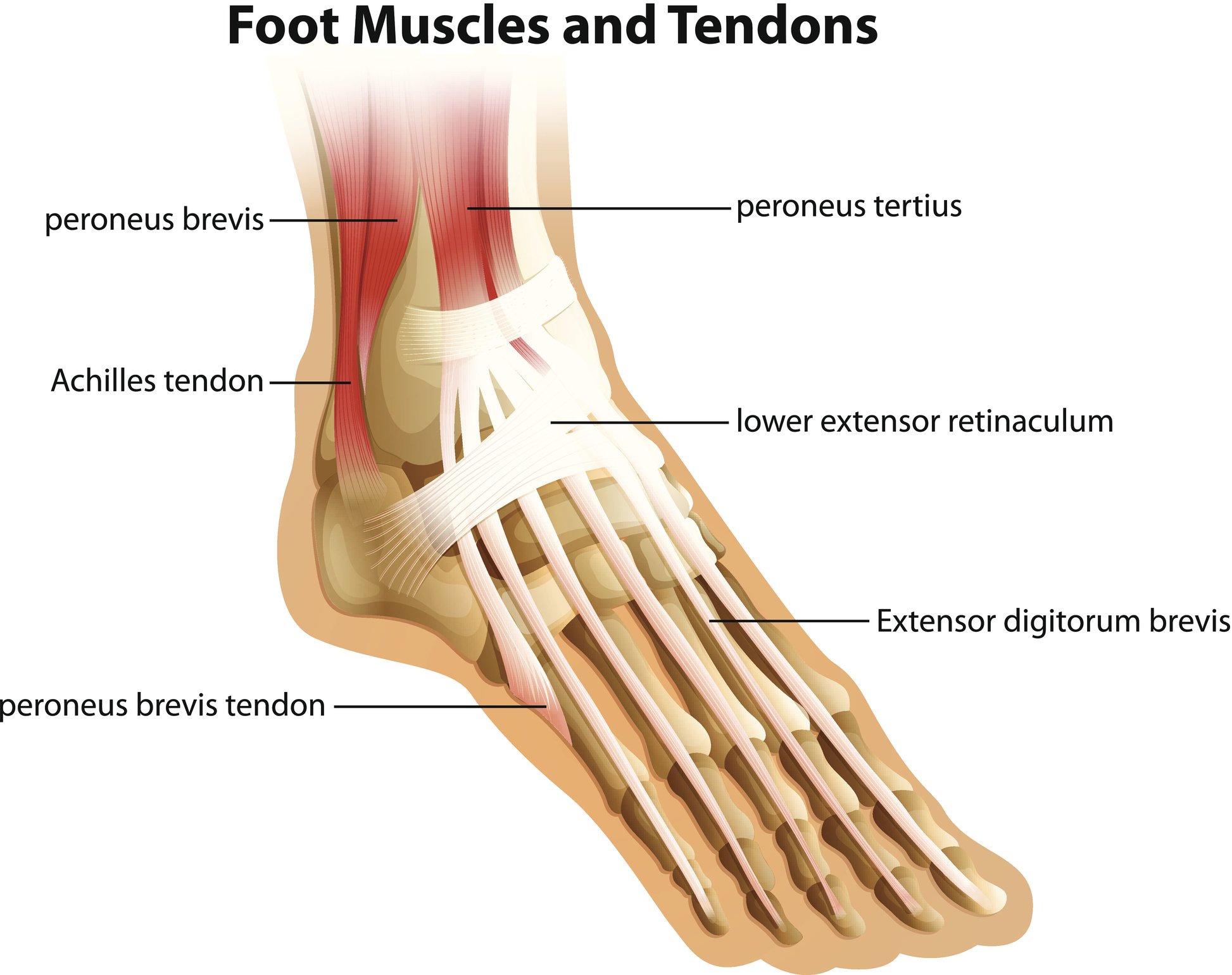
The tibilias anterior which allows the foot to move upward. The arches of the foot formed by the tarsal and metatarsal bones strengthened by ligaments and tendons allow the foot to support the weight of the body in the erect posture with the least weight. 291 page 277 is composed of the calcaneus the cuboid.
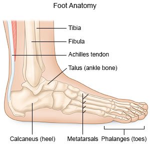
The bones of the foot are organized into rows named tarsal bones metatarsal bones and phalanges. The talus bone supports the leg bones tibia and fibula forming the ankle. The lateral arch see fig.
These bones are arranged into longitudinal and transverse arches with the support of various muscles and ligaments. Arches of the foot. The metatarsal arch is the primary transverse arch running across the foot just behind the ball.
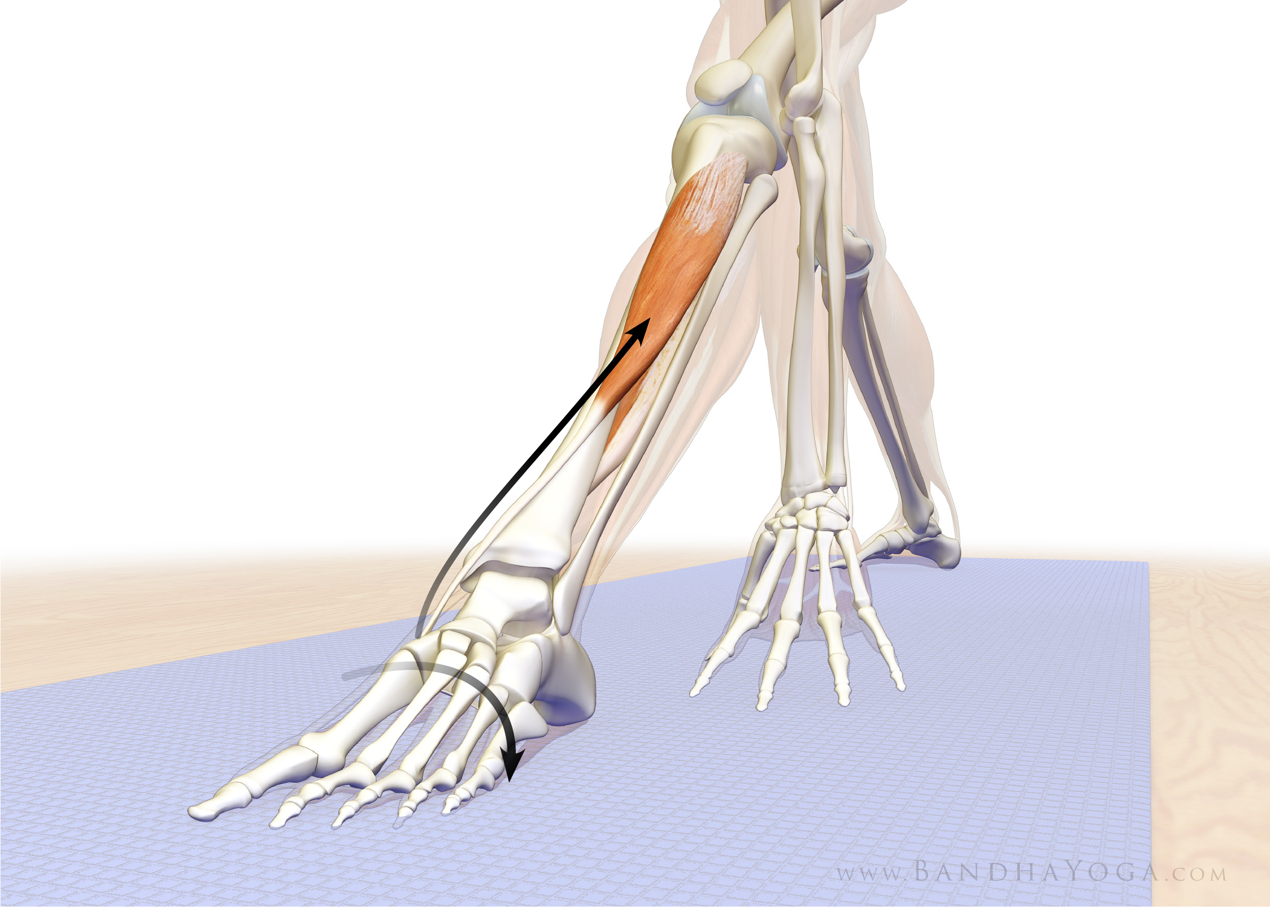
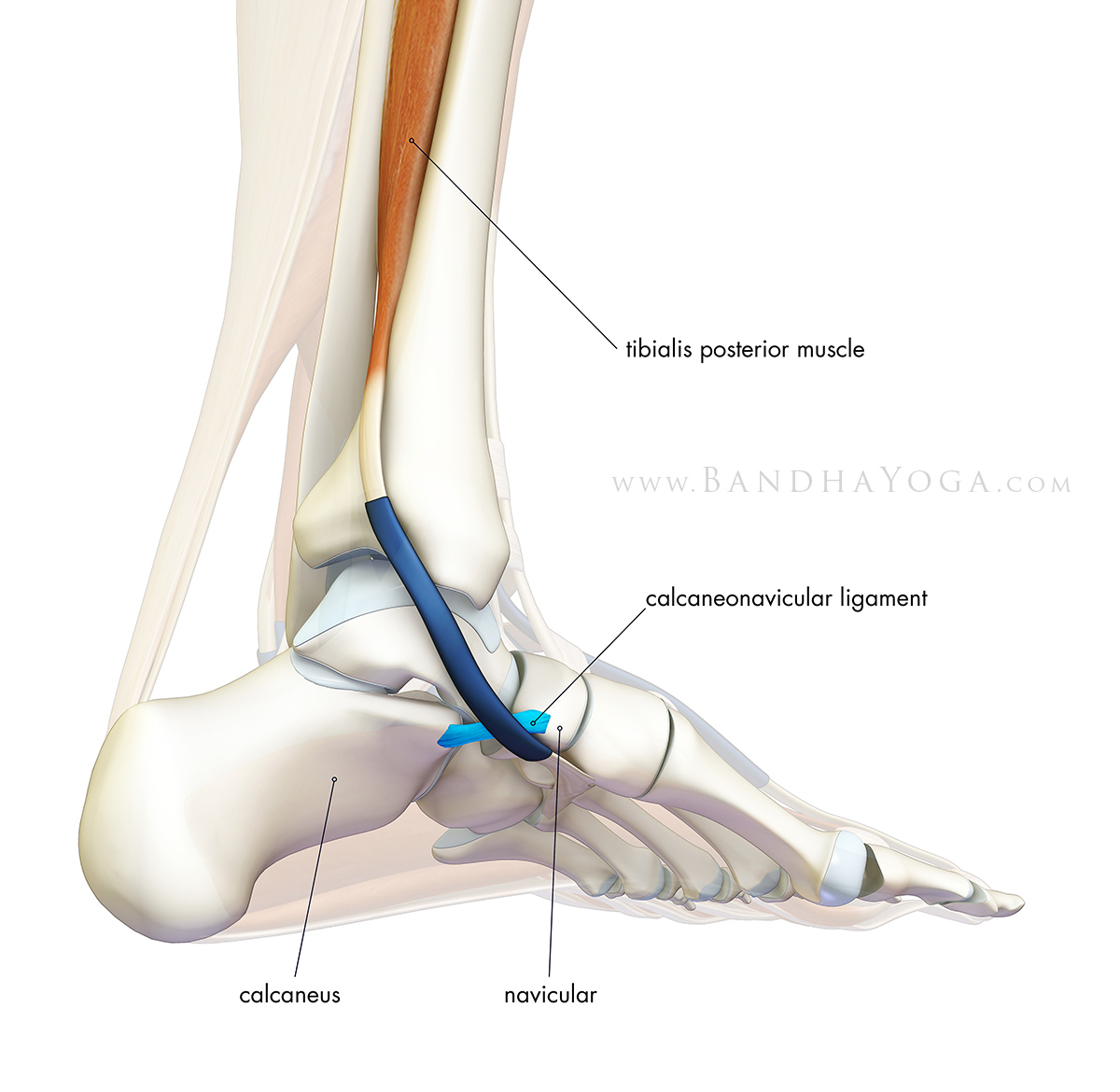
The tibilias posterior which supports the foots arch. The arch is further supported by the plantar aponeurosis by the small muscles in the sole of the foot by the tendons of the tibialis anterior and posterior and peronæus longus and by the ligaments of all the articulations involved. The hindfoot forms the heel and ankle.
Foot definition the foot is a part of vertebrate anatomy which serves the purpose of supporting the animals weight and allowing for locomotion on land. Twenty muscles give the foot its shape support and the ability to move.
 The Foot Advanced Anatomy 2nd Ed
 The Daily Bandha Trikonasana Part Three Foot Arch Mastery
 Amazon Com Yxfyxf Human Normal Foot Flat Foot High Arch
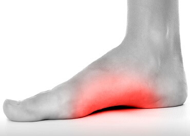 Foot Arch Pain Causes Treatment Foot Pain Explored
 Arches Of The Foot Physiopedia
 Foot Arch Pain Plantar Fasciitis Fallen Arches Flat Feet
 4 Ways To Prevent And Treat Posterior Tibial Tendonitis
 Foot Arch Pain

 A Closer Look At The Arches Of The Foot Mass4d Foot Orthotics
The Longitudinal Arches Of The Feet In Yoga
 Foot Arch Pain Causes Treatment Foot Pain Explored
 Foot Anatomy Bones Ligaments Muscles Tendons Arches
 Plantar Fasciitis Causes Symptoms And Treatment Info
 High Arches Problems How To Relieve Foot Arch Pain
 Exercises For Plantar Fasciitis
 Arch Strain What Causes Pain In The Arch Of The Foot
 Foot Anatomy Animated Tutorial
 Different Foot Arch Types And What It Means For You Feet
 Anatomy Arches Of Foot By Geeta Goswami
 Foot Pain Diagnosis Achilles Tendinitis Causes Home
 Easy Notes On Arches Of The Foot Learn In Just 3 Minutes
 Muscles That Lift The Arches Of The Feet
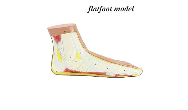
 Flatfoot What You Need To Know
Belum ada Komentar untuk "Anatomy Of The Foot Arch"
Posting Komentar