Aorta Branches Anatomy
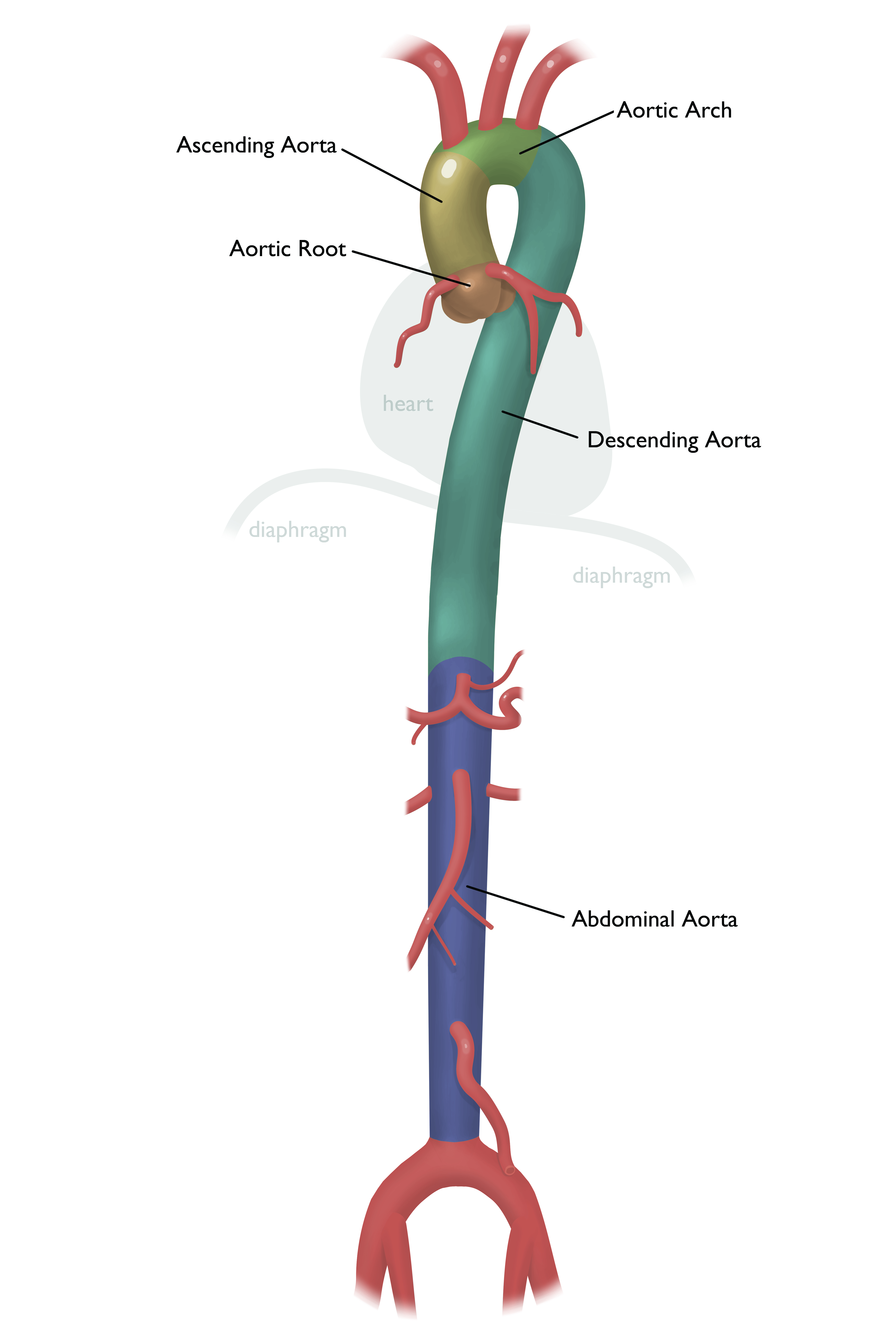
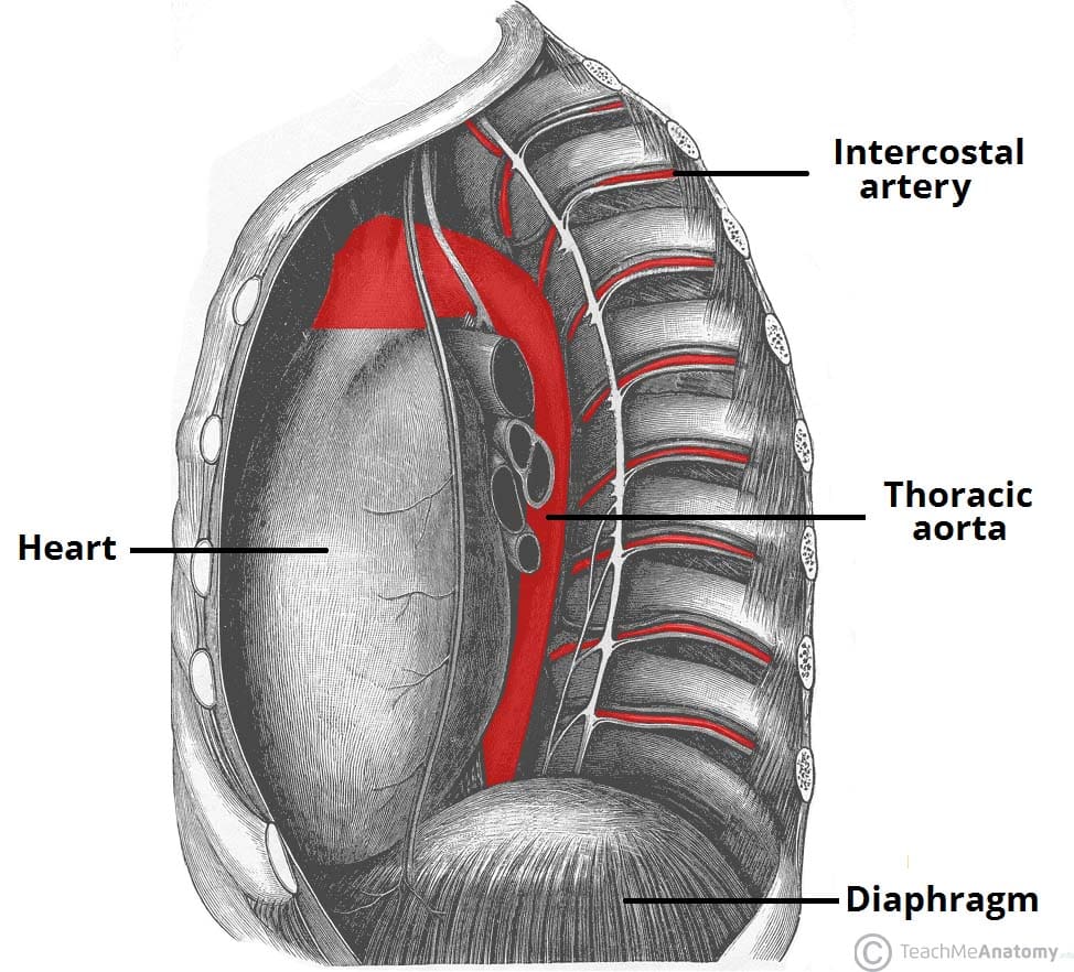
The descending aorta runs down through the posterior centre of the trunk past the heart lungs and esophagus through an opening in the diaphragm and into the abdominal cavity. Eɪ ˈ ɔːr t ɪ k is the part of the aorta between the ascending and descending aorta.
 Illustration Of Aorta And Branches Stock Image C017 2678
Descending thoracic aorta pericardial branches.

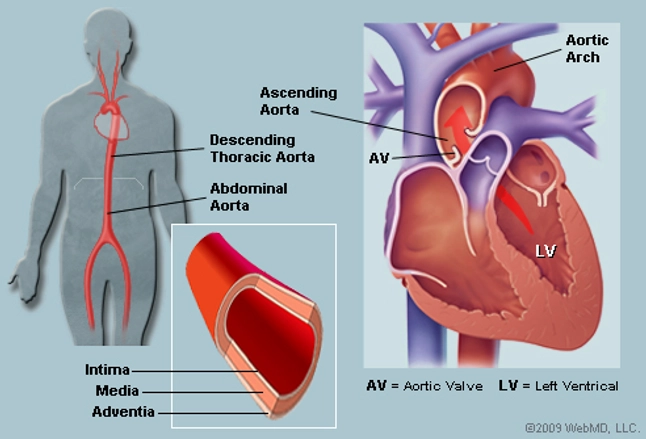
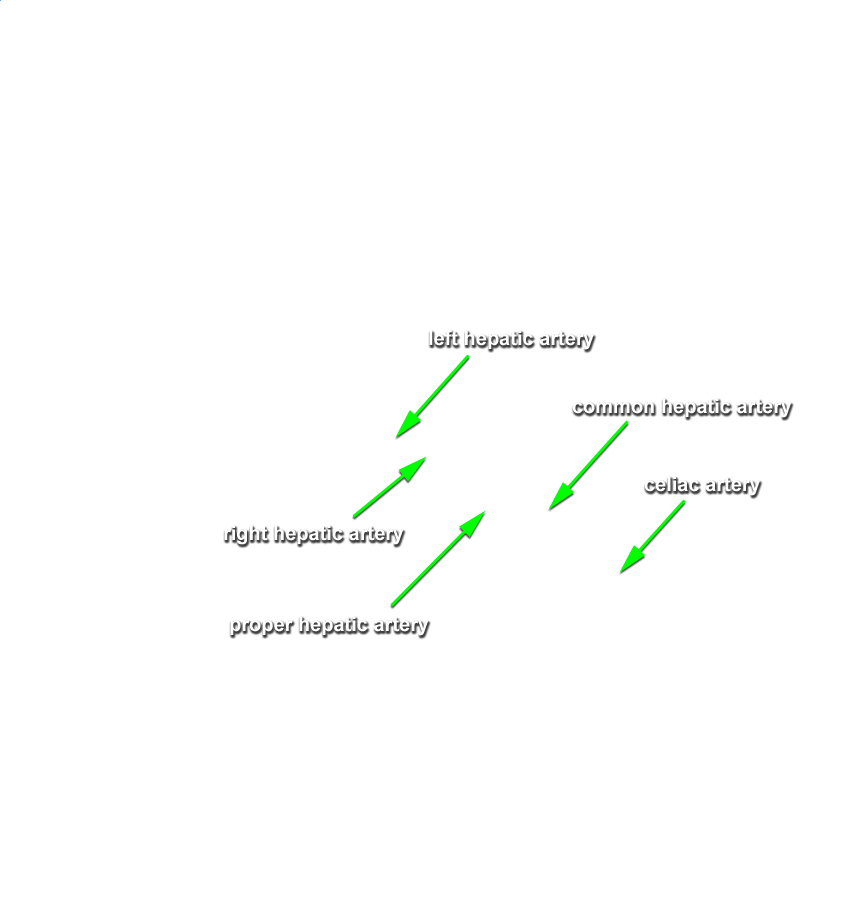
Aorta branches anatomy. Lymph nodes nerves vessels areolar tissue of the posterior mediastinum. Picture of the aorta the ascending aorta rises up from the heart and is about 2 inches long. It is 2 inches long in length and travels with the pulmonary trunk in the pericardial sheath.
The left and right aortic sinuses are dilations in the ascending aorta located at the level of the aortic valve. The aortic arch arch of the aorta or transverse aortic arch english. 3 single anterior visceral branches coeliac superior mesenteric artery inferior mesenteric artery.
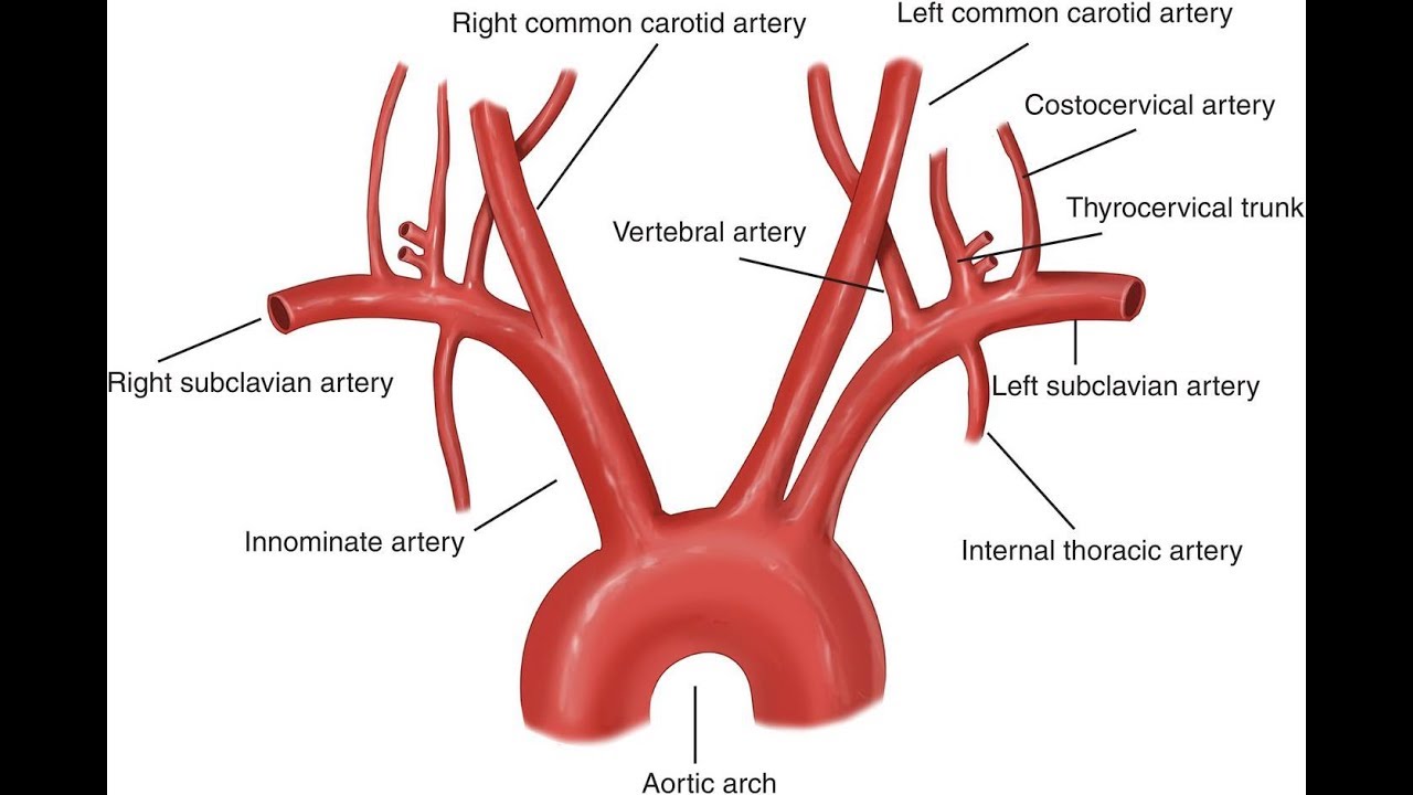
The three main arteries branch from the aortic arch and give rise to further branches that supply oxygenated blood to the head neck upper limbs and upper part of the body. The arch travels backward so that it ultimately runs to the left of the trachea. The only branches of the ascending aorta are the right and left coronary arteries that supply the myocardium of the heart.
The ascending aorta arises from the aortic orifice from the left ventricle and ascends to become the aortic arch. The abdominal aorta begins at. The descending thoracic aorta travels down through the chest.
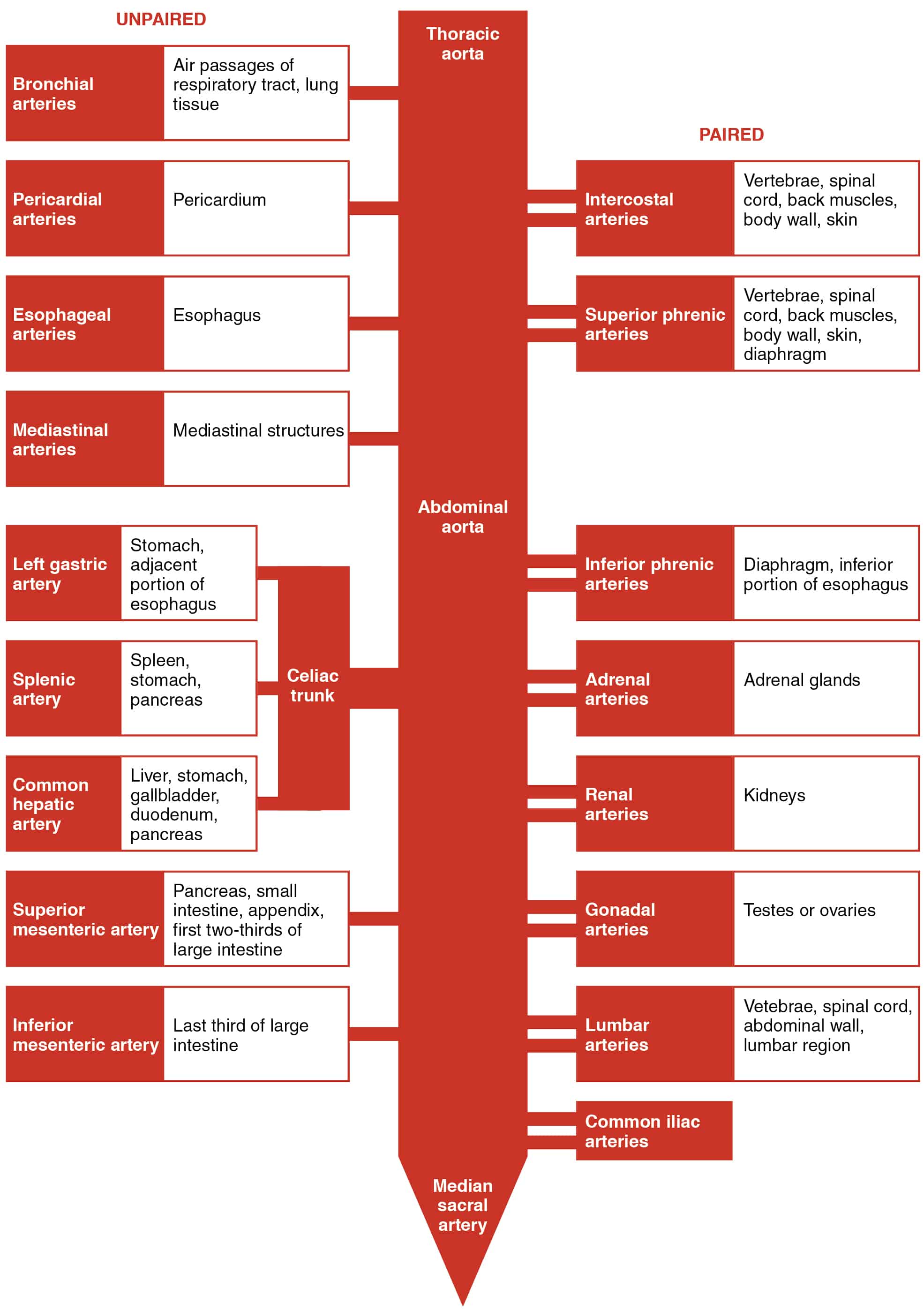
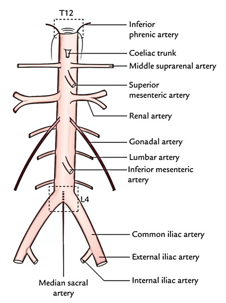
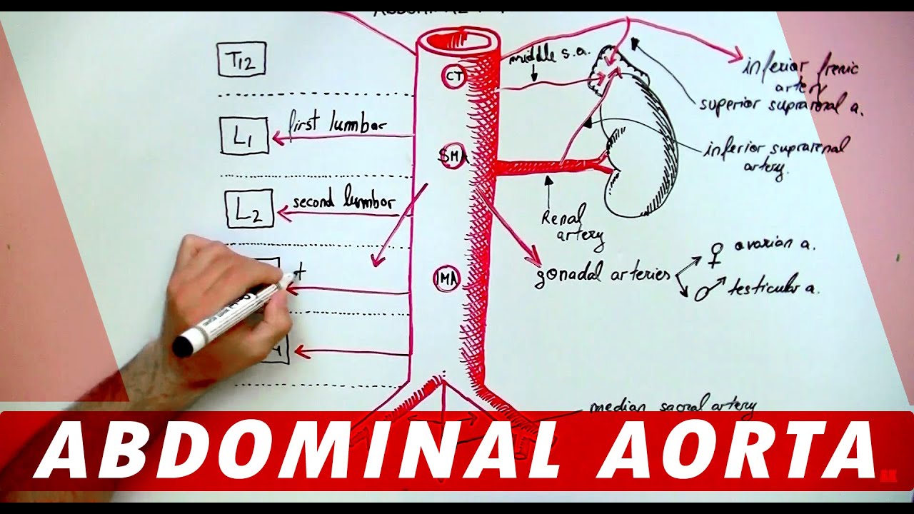
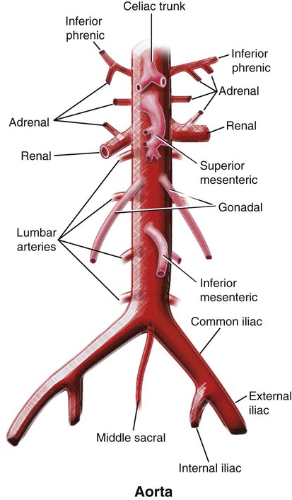
The abdominal aorta has. 3 paired lateral visceral branches suprarenal renal gonadal. Bronchial tree below the trachea.
The aortic arch curves over the heart giving rise to branches that bring blood to the head neck. Then the ascending aorta arches to the left forming the arch of the aorta which descends and ends at the level of the intervertebral disc between the fourth and fifth thoracic vertebrae. 3 terminal branches two.
The branches of the thoracic aorta run to various organs esophagus lungs adrenal glands and tissues pericardium diaphragm abdominal wall muscles posterior intercostal spaces in order to. Posterior aspect of the pericardial sac. 5 paired lateral abdominal wall branches inferior phrenic and four lumbar.
 The Aorta Branches Aortic Arch Teachmeanatomy
 Chapter 22 Diseases Of The Aorta Multimodal
 The Aorta Human Anatomy Picture Function Location And
 Aorta Anatomy Uf Health Aortic Disease Center Overview
 Aortic Arch Human Anatomy Organs
![]() Aorta Anatomy Branches Course Divisions Kenhub
Aorta Anatomy Branches Course Divisions Kenhub
 Branches Of The Aorta Abdominal Region Human Anatomy Organs
 Branches Of The Aorta Thoracic Section Human Anatomy Organs
Coronary Circulation Wikipedia
 Print Anatomy Of The Arteries Of The Trunk Lecture 5
 Circulatory Routes Boundless Anatomy And Physiology
 Branches Of The Aorta Abdominal Region Human Anatomy
 Anatomy Pathways Thoracic And Abdominal Vasculature
 Abdominal Aorta Anatomy Of The Abdomen
Human Body Anatomy Thoracic Aorta And Major Branches By
 Easy Notes On Abdominal Aorta Learn In Just 3 Minutes
 Abdominal Aorta And It S Branches Anatomy
 Anatomy Abdominal Aorta Branches
 The Aorta Branches Aortic Arch Teachmeanatomy
 Branches Of Abdominal Aorta Anatomy Lecture For Medical Students Usmle Step 1
 Vascular Anatomy Of The Pelvis Radiology Key
 Anatomy Lab 13 Branches Of Abdominal Aorta Diagram Quizlet
 The Big Vessels Cardiovascular System
 Aorta And Its Branches Anatomy
 Image Result For Abdominal Aorta Branches Abdominal Aorta


Belum ada Komentar untuk "Aorta Branches Anatomy"
Posting Komentar