Foot Anatomy Xray
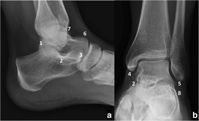
The distal tibia and fibula articulate with each other at the distal tibiofibular joint which is more commonly referred to as the tibiofibular syndesmosis or simply the syndesmosis. Osseous radiographic anatomy of the upper extremity duration.
 Optima Xr646 Hd X Ray System Powered By Helix Ge Healthcare
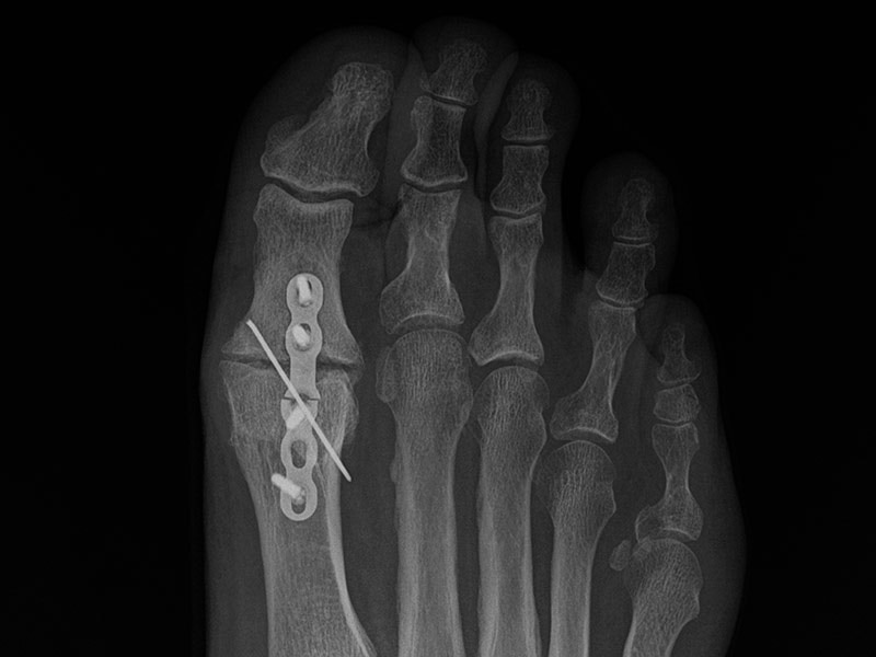
Head of the 1st metatarsal.
Foot anatomy xray. This wallpaper was upload at january 21 2018 upload by admin in anatomy diagram. You can download foot anatomy xray in your computer by clicking resolution image in download by size. Ankle is joint that is located between leg and foot a main contributor of stability sunday december 15 2019.
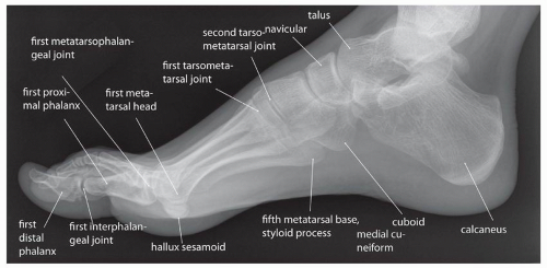
Remember to check the whole film though. Normal right foot radiographs in a young adult female for reference. Foot radiograph an approach foot radiographs are commonly performed in emergency departments usually after sport related trauma and often with a clinical request that states lateral border pain.
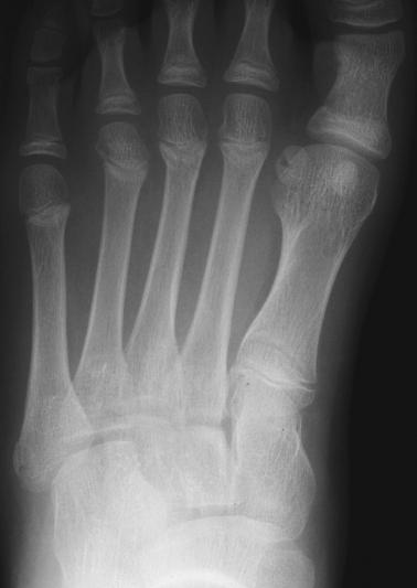
The foot series is comprised of a dorsoplantar dp medial oblique and a lateral projection. 1 fibula 2 cuboid 3 5th metatarsal bone 4 tibia 5 talus 6 navicular 7 cuneiform 8 1st metatarsal bone 9 proximal phalanx 10 distal phalanx. Xray medial oblique foot anatomy.
Often a foot x ray is also requested for the investigation of osteomyelitis arthritides or. Douglas gillard bs dc spine researcher 17151 views. 1 calcaneus 2 cuboid 3 5th metatarsal bone 4 talus 5 navicular 6 cuneiform.
Ip interplangeal joint. Dont forget to rate and comment if you interest with this wallpaper. Anatomy the ankle is a synovial joint composed of the distal tibia and fibula as they articulate with the talus.
When checking any post traumatic foot x ray it is crucial to assess alignment of the bones at the joints. Proximal phalanx of the 1st digit. Ankle anatomy sprain clinical anatomy fracture radiology x ray.
Approach to foot series. This webpage presents the anatomical structures found on foot radiograph. Loss of joint alignment can represent severe injury even in the absence of a fracture.
The series is often utilized in emergency departments after trauma or sports related injuries 24. Foot anatomy xray is free hd wallpaper.
 Equine Podiatry Dr Stephen O Grady Veterinarians
Rheumatoid Arthritis Of The Foot And Ankle Orthoinfo Aaos
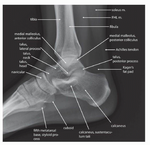
 Normal Radiographic Anatomy Of The Ankle Radiology Case

Royalty Free Foot Xray Stock Images Photos Vectors
 Royalty Free Foot Xray Stock Images Photos Vectors
 X Enkel Startradiology
 Foot X Rays
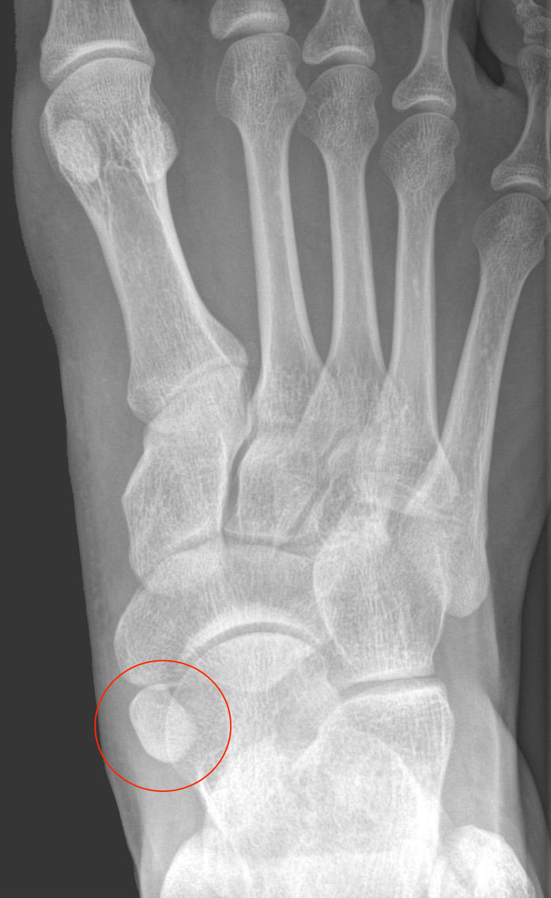
 Foot Annotated X Ray Radiology Case Radiopaedia Org
 Radiology In Ped Emerg Med Vol 4 Case 14

 Cone Beam Computed Tomography With Load Technique Wbct
 Anatomical Variation In The Ankle And Foot From Incidental
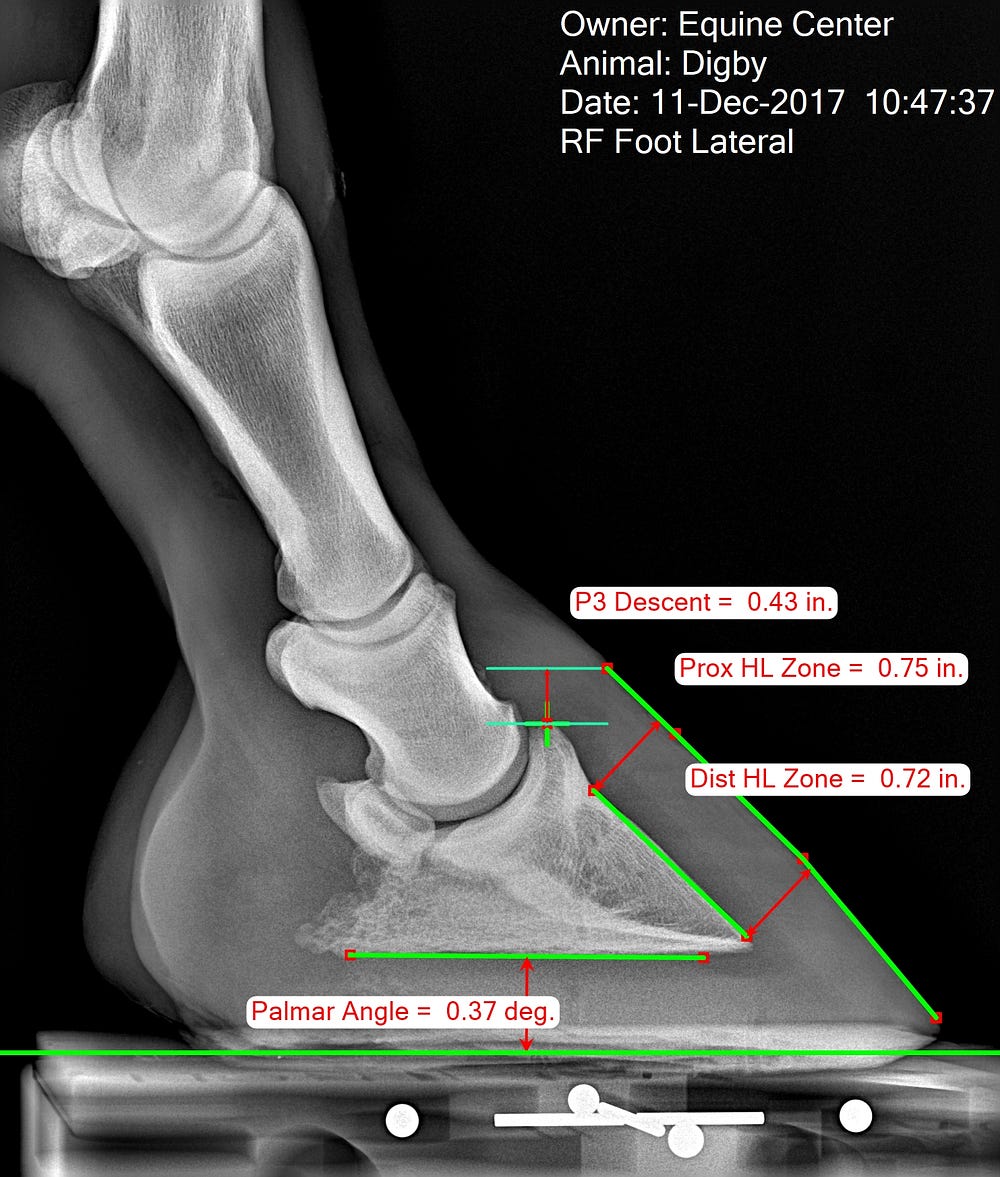 Measuring The Equine Hoof In Radiographs Mc Ai

 852 Calcaneus And Foot Anatomy The Xray Shows A Lateral
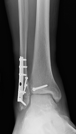 Broken Ankle Types Of Fractures Diagnosis Treatments
 Diagnostic Imaging Techniques Of The Foot And Ankle
 Pediatric Ankle And Foot Injuries Sciencedirect

 Oblique And Anterior Posterior View X Rays Of A Normal Foot
 Pin By Jennifer Endres Serrao On X Ray Foot Anatomy
 Basketball Player With Left Foot Pain
 Radiology In Ped Emerg Med Vol 4 Case 14
 Diagnostic Imaging Techniques Of The Foot And Ankle
 A Lateral And B Oblique Foot Radiographs Of The Patient
Belum ada Komentar untuk "Foot Anatomy Xray"
Posting Komentar