Zygomatic Arch Anatomy
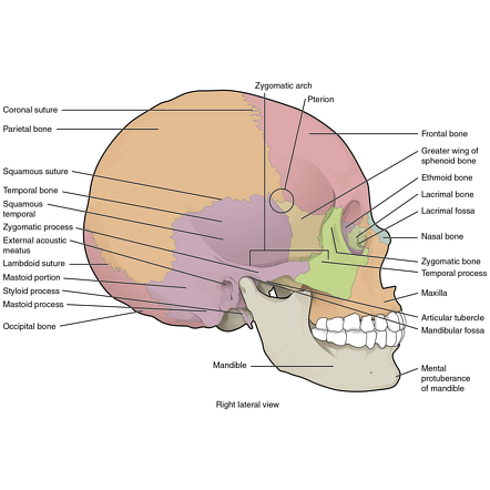
A posterior which runs backward above the external acoustic meatus and is continuous with the supramastoid crest. The zygomatic bone is small and quadrangular and is situated at the upper and lateral part of the face.
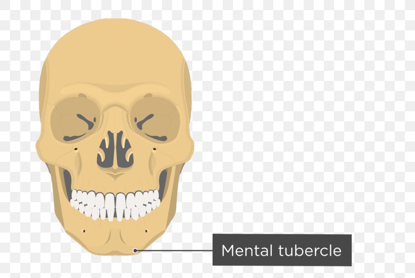
 The Zygomatic Bone Human Anatomy
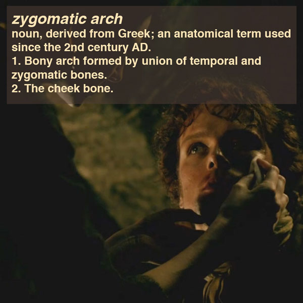
The bony arch at the outer border of the eye socket formed by the union of the cheekbone and the zygomatic process of the temporal bone.
Zygomatic arch anatomy. Myofascial trigger point treatment for headache and tmd. Musculoskeletal and neurologic diseases. Surgery of the orbit.
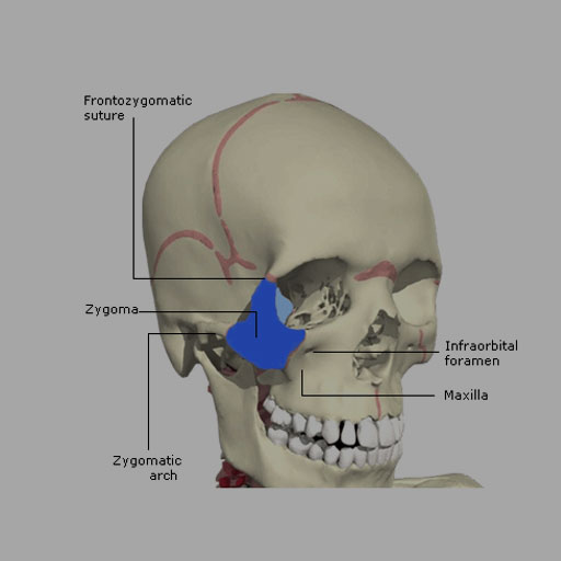
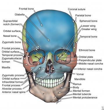
It adjoins the frontal bone at the outer edge of the orbit and the sphenoid and maxilla within the orbit. The zygomatic arch is formed by the union of the temporal process of the zygomatic bone and the zygomatic process of the temporal bone at the zygomaticotemporal suture. The masseter muscle important in chewing arises from the lower edge of the arch.
Disorders of the eye and vision. The zygomatic bone forms in membrane ie. Zygomatic process of the temporal bone linked by the temporozygomatic suture.
Frontal bone via the frontozygomatic suture which creates the rounded form of the bony orbit. The zygomatic process of the temporal arises by two roots. Each zygomatic bone articulates with the temporal bone frontal bone maxilla and sphenoid bones.
Le fort type 3 fracture. Introduction to temporal bone anatomy. Zygomatic arch noun anatomy.
Another major chewing muscle the temporalis passes through the arch. Several bones and joints surround the zygoma including the. Zygomatic arch temporomandibular joint dysplasia.
Zygomatic arch bridge of bone extending from the temporal bone at the side of the head around to the maxilla upper jawbone in front and including the zygomatic cheek bone as a major portion. An anterior directed inward in front of the mandibular fossa where it expands to form the articular tubercle. The anatomy of the common marmoset.
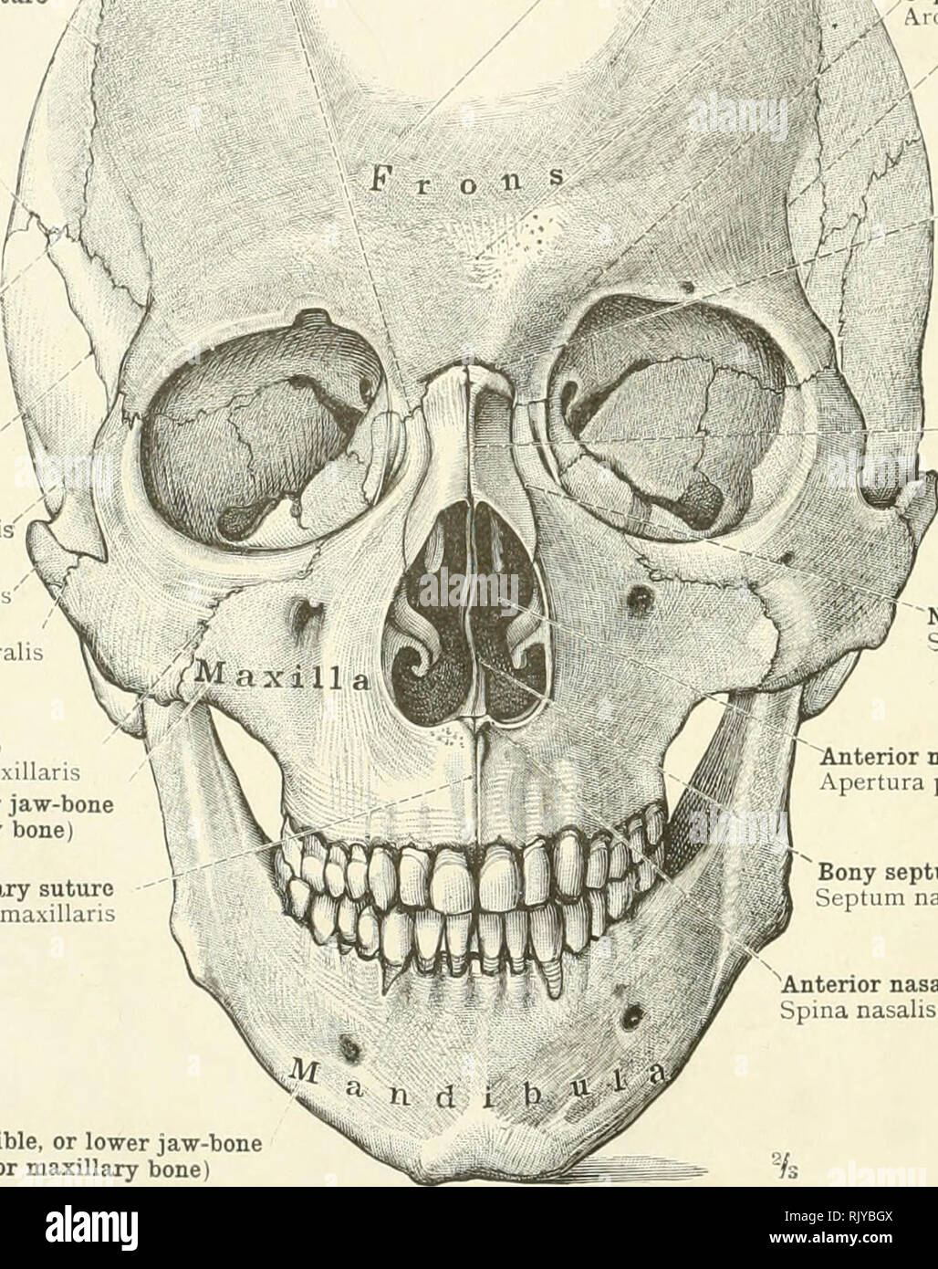
They are also commonly referred to a as the cheekbones or malar bones l mala the cheek. The zygomatic bones gr zygoma yoke are two facial bones that form the cheeks and the lateral walls of the orbits. Zygomatic process of the maxillary bone articulated by the.
It forms the central part of the zygomatic arch by its attachments to the maxilla in front and to the zygomatic process of the temporal bone at the side. It forms the prominence of the cheek part of the lateral wall and floor of the orbit and parts of the temporal and infratemporal fossæ fig.
 Zygomatic Arch Archives Md Direct
 Zygomatic Bone Zygomatic Process Of Temporal Bone Zygomatic
![]() Zygomatic Bone Anatomy And Pathology Kenhub
Zygomatic Bone Anatomy And Pathology Kenhub
 Anp 1107 Lecture Notes Winter 2017 Lecture 3 Zygomatic
 Zygomatic Process Wikipedia
 Zygomatic Process Of Temporal Bone Zygomatic Bone Zygomatic
 Anatomical Location Of The Apg Region Green Area Yellow D
 Zygomatic And Nasal Injury Rcemlearning
 Skeletons And Skulls Mandlble Zygomatic Arch Temporal Bone
 Zygomatic Arch Zygoma Cheek Bone Definition Quiz
 Zygomatic Arch Radiology Reference Article Radiopaedia Org
 Ce4rt X Ray Positioning Guide For Radiologic Techs
 4 1 Diagram Zygomatic Arch Diagram Quizlet
 Ancestral Variations In The Shape And Size Of The Zygoma
 Zygomatic Arch
 Midface Reduction Fixation Orif 4 Point Fixation
 Ontogeny Of Bone Strain The Zygomatic Arch In Pigs
 An Atlas Of Human Anatomy For Students And Physicians
 Anatomy Temporal Fossa And Zygomatic Arch At University Of

 Facial Bone Anatomy Overview Mandible Maxilla
 Zygomatic Arch An Overview Sciencedirect Topics
 Ce4rt X Ray Positioning Guide For Radiologic Techs
 Zygomatic Arch Outlander Anatomy
 The Zygomatic Bone Human Anatomy
Zygomatic Arch
 Zygomatic Arch Stock Photos Zygomatic Arch Stock Images
 A Caa In A Non Syndromic Patient With Complete Preservation
 Zygomatic Arch An Overview Sciencedirect Topics
Belum ada Komentar untuk "Zygomatic Arch Anatomy"
Posting Komentar