Anatomy Of Acl
The main part of the anterior cruciate ligament consists of type i collagen positive dense connective tissue. The anterior cruciate ligament acl is one of a pair of ligaments in the center of the knee joint that form a cross and this is where the name cruciate comes from.
Anterior Cruciate Ligament Acl Injuries Orthoinfo Aaos
It also it stabilizes the knee during all types of movements.

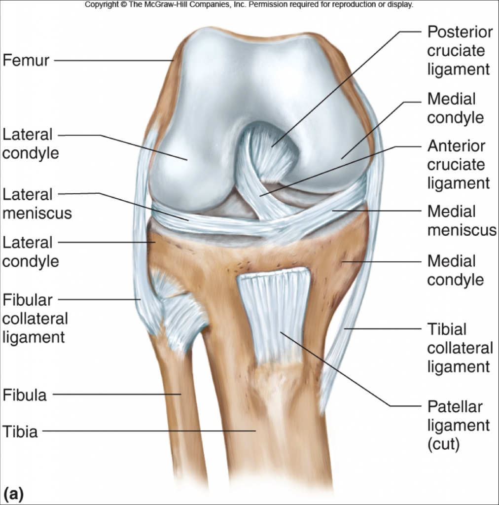
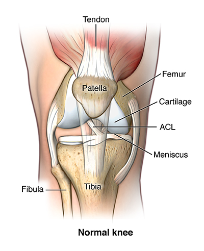
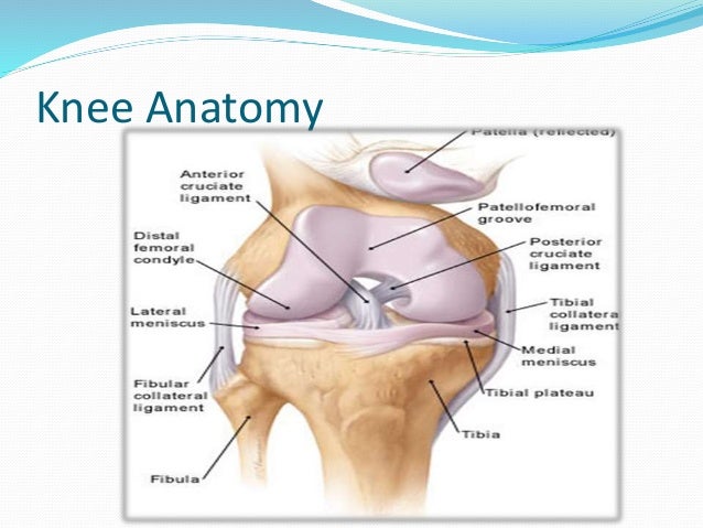
Anatomy of acl. When working with any injury it is important to note. The acl originates from deep within the notch of the distal femur. In terms you may have heard before the acl connects the thigh bone to the shin bone.
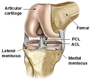
The longitudinal fibrils of type i collagen are divided into small bundles by thin type iii collagen positive fibrils. Anterior cruciate ligament anatomy anterior cruciate ligament function during movement of the knee the anterior cruciate ligament acl prevents anterior sliding of the tibia. The posterior cruciate ligament prevents posterior sliding of the tibia.
Anatomy of an injury. Its proximal fibers fan out along the medial wall of the lateral femoral condyle. The acl is a key structure in the knee joint as it resists anterior tibial translation and rotational loads.
The acl controls anterior movement of the tibia and inhibits extreme ranges of tibial rotation. In the distal third the structure of the tissue varies from the typical structure of a ligament. There is both an anterior cruciate ligament acl and a posterior cruciate ligament pcl.
29th annual mizzou nsslha update seminar. The anterior cruciate ligament acl is a band of dense connective tissue which courses from the femur to the tibia. The anatomy of the anterior cruciate ligament acl.
The anterior cruciate ligament acl the anterior cruciate ligament acl. Anterior cruciate ligament acl is one of the two cruciate ligaments that stabilize the knee joint. The anterior cruciate ligament acl is one of a pair of cruciate ligaments the other being the posterior cruciate ligament in the human knee.
The 2 ligaments are also called cruciform ligaments as they are arranged in a crossed formation. The component acl bundles are named based on their tibial insertion. Anterior cruciate ligament anatomy.
The majority of authorities believe that the acl consists of 2 major bundles the posterolateral bundle pl and the anteromedial bundle am. The mizzou chapter of the national student speech language hearing association is proud to host our 29th update seminar and we warmly welcome invited speaker. Gross anatomy the acl arises from the anteromedial aspect of the intercondylar area on the tibial plateau and passes upwards and backwards to.
 Acl Tears Pinnacle Orthopaedics
Common Knee Injuries Orthoinfo Aaos
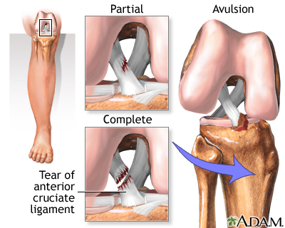 Anterior Cruciate Ligament Acl Injury Medlineplus Medical
The Anterior Cruciate Ligament Acl
 Anterior Cruciate Ligament Acl Injury Orthopedics
 Anterior Cruciate Ligament Wikipedia
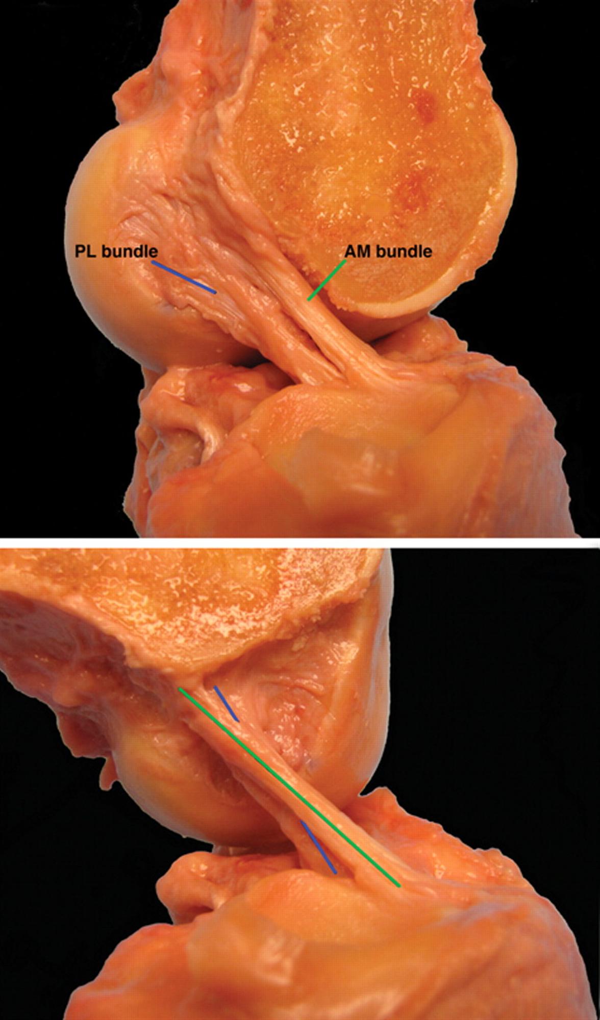 Ligaments Of The Knee Knee Sports Orthobullets
 Anterior Cruciate Ligament Acl Tears
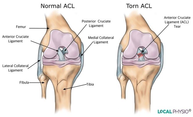 Anterior Cruciate Ligament Acl Injury Local Physio
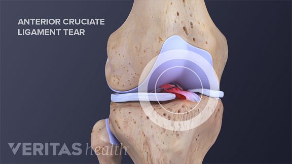 Anterior Cruciate Ligament Acl Tears
Anatomic Double Bundle Anterior Cruciate Ligament
 Knee Human Anatomy Function Parts Conditions Treatments
 Knee Injuries Flashcards Quizlet
 The Knee Resource Acl Anterior Cruciate Ligament Rupture
 Anterior Cruciate Ligament Acl
 Normal Acl Pcl Left Knee Anatomy Ruptured Acl Mri Of
 Anterior Cruciate Ligament Acl Injuries Core Em
 Biomechanics Functional Anatomy Behind Acl Tears Athletix
Acl Regeneration Promising Two Year Results Of First In
 Anterior Cruciate Ligament Injury Diagnosis Management
 Anatomy And Epidemiology Of Acl Injury
Anterior Cruciate Ligament Acl Injuries Orthoinfo Aaos
 Care Of Your Knee Following Anterior Cruciate Ligament Acl

Belum ada Komentar untuk "Anatomy Of Acl"
Posting Komentar