Anatomy Of The Mandible
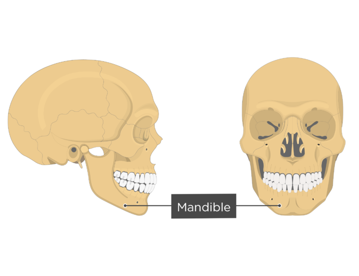
It is the moving part of the jaws when the body is engaged in the feeding process and for that reason all the muscles of mastication including the medial and lateral pterygoid muscles the temporal muscle and the masseter muscle attach to it. Two vertical portions form movable hinge joints on either side of the head articulating with the glenoid cavity of the temporal bone of the skull.
 Anatomy Of The Mandible And The Ian In Relation To The
Learn all about its anatomy and vascular supply at kenhub.
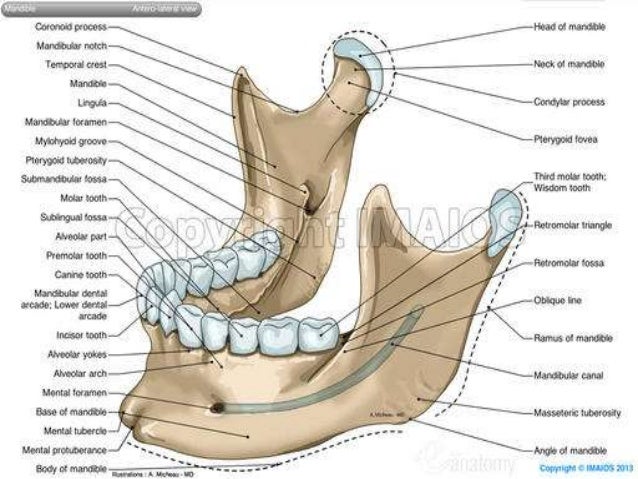
Anatomy of the mandible. Learn all about it at kenhub. This feature is not available right now. Dysfunction of the tmj can cause severe pain and lifestyle limitation.
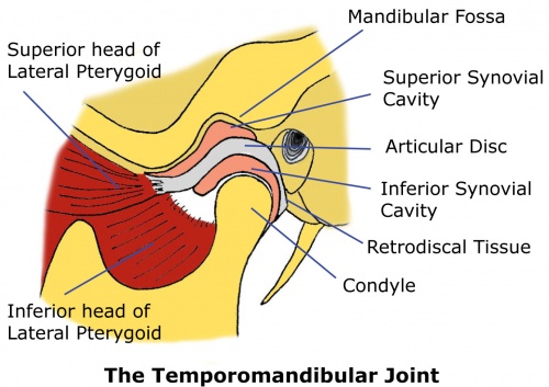
Tmj anatomy the temporomandibular joint tmj or jaw joint is a bi arthroidal hinge joint that allows the complex movements necessary for eating swallowing talking and yawning. The mandible is a u shaped bone. Please try again later.
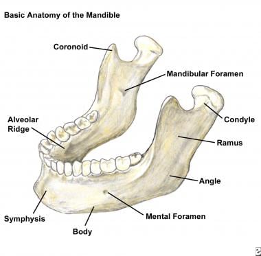
The mandible consists of a horizontal arch which holds the teeth and contains blood vessels and nerves. The temporomandibular joint connects the skull with the lower jaw. The body found at the front.
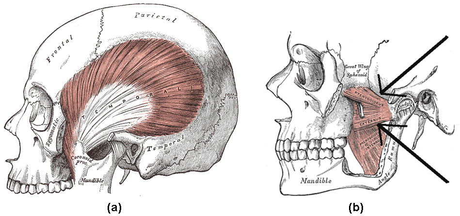
These muscles are the masseter the temporalis the medial pterygoid and the lateral pterygoid. Fractures of the neck of the mandible are often transverse and usually accompanied. The mandible is composed of 2 hemimandibles joined at the midline by a vertical symphysis.
Fractures of the coronoid process are uncommon and usually singular. Fractures of the angle of the mandible are usually oblique and may involve the. The mandible is a singular bone that has a distinctive horse shoe shape and is symmetrical on both sides.
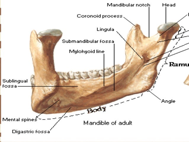
The muscles work in combination to pivot the lower jaw up and down and to allow movement of the jaw from side to side. The characteristics of mandibular fractures are as follows. A ramus on the left and the right the rami rise up from the body of the mandible and meet with the body at the angle of the mandible or the gonial angle.
It is the only mobile bone of the facial skeleton and since it houses the lower teeth its motion is essential for mastication. The rami also provide attachment for muscles important in chewing. Each of these muscles occurs in pairs with one of each muscle appearing on either side of the skull.
Masseter muscle this article describes the anatomy origin insertion innervation blood supply and function of the masseter muscle. It is formed by intramembranous ossification. It consists of a curved horizontal portion the body and two perpendicular portions the rami which unite with the ends of the body nearly at right angles.
The mandible the largest and strongest bone of the face serves for the reception of the lower teeth. The mandible consists of.
Dental Malpractice Central Anatomy Of The Lingual Nerve
 The Mandible Lower Jaw Human Anatomy
 Pin By Renee Mccarty On Diagnostic Imaging Dental Anatomy
 Male Mandible Bone Skull Anatomy Isolated On White
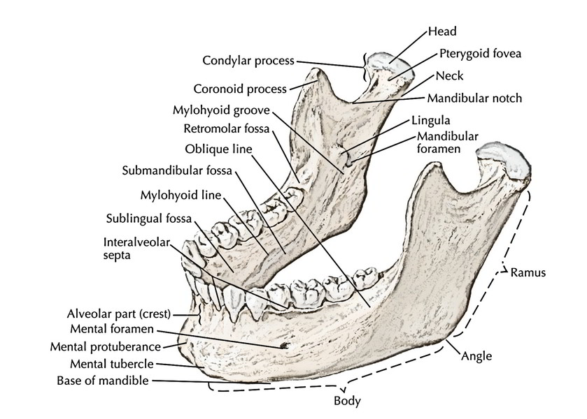 Easy Notes On Mandible Learn In Just 4 Minutes Earth S Lab
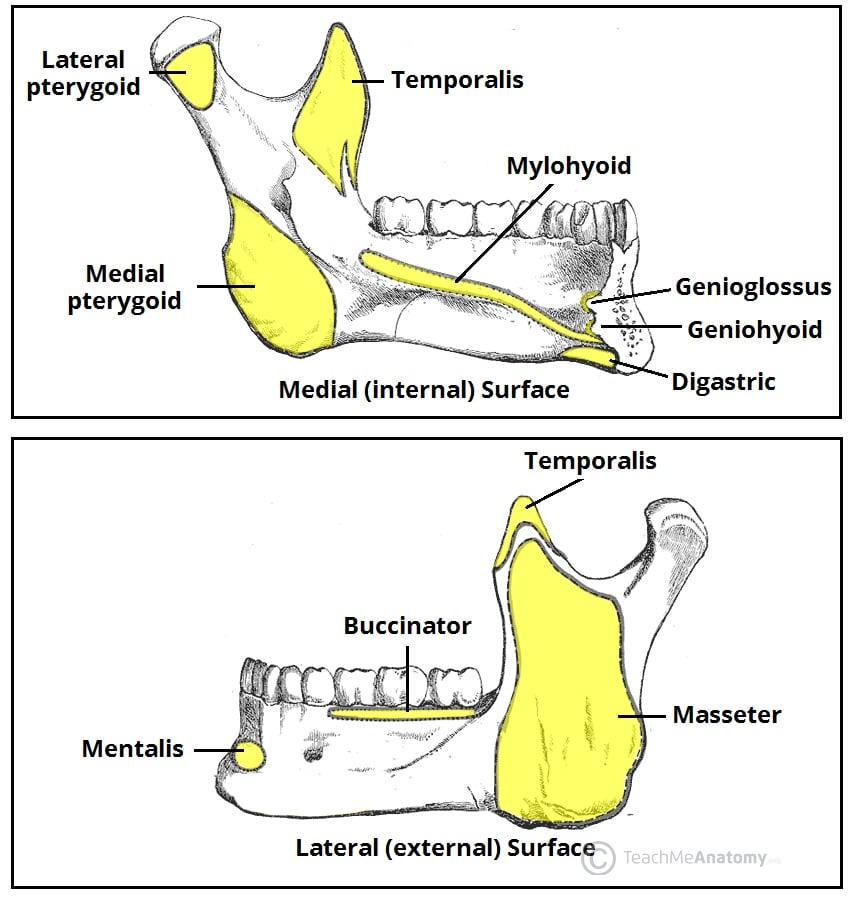 The Mandible Structure Attachments Fractures
 Give Mental Block In The Mental Foramen Mandible Mandible
 Human Skull Replica Life Size Articulating Mandible Jaw
 Anatomy Of Maxilla And Mandible
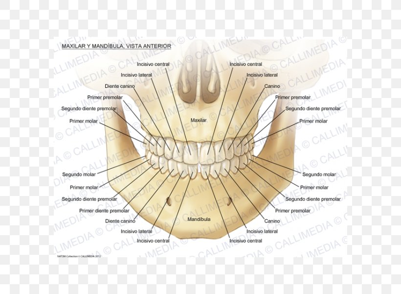 Maxilla Anatomy Mandible Mandibular Nerve Human Body Png
 Mandible Radiology Reference Article Radiopaedia Org
:watermark(/images/watermark_only.png,0,0,0):watermark(/images/logo_url.png,-10,-10,0):format(jpeg)/images/anatomy_term/mandible-bone/5JWY3peYMVzslzYGo0eFw_MandibleBone.png) The Mandible Anatomy Structures Fractures Kenhub
The Mandible Anatomy Structures Fractures Kenhub
 Mandibular Fracture Imaging Practice Essentials
 Mandibular Nerve An Overview Sciencedirect Topics
 Articulation Of The Mandible Human Anatomy
 The Mandible Anatomy Images Illustrations Anatomy Images
 Mandible Download Free 3d Model By University Of Dundee
 Schema Showing The Gross Anatomy Of The Mandible Download
 Head And Neck Muscles Boundless Anatomy And Physiology
The Anatomy Of The Laboratory Mouse
 Anatomy Of Oromandibular Cancer Headandneckcancerguide Org
:watermark(/images/watermark_5000_10percent.png,0,0,0):watermark(/images/logo_url.png,-10,-10,0):format(jpeg)/images/atlas_overview_image/552/W39arzeteG61UaMYzqsYw_skull-anterior-lateral-views_english.jpg) The Mandible Anatomy Structures Fractures Kenhub
The Mandible Anatomy Structures Fractures Kenhub

Belum ada Komentar untuk "Anatomy Of The Mandible"
Posting Komentar