Anatomy Hamstring
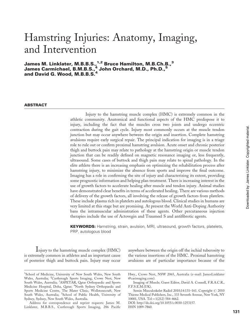
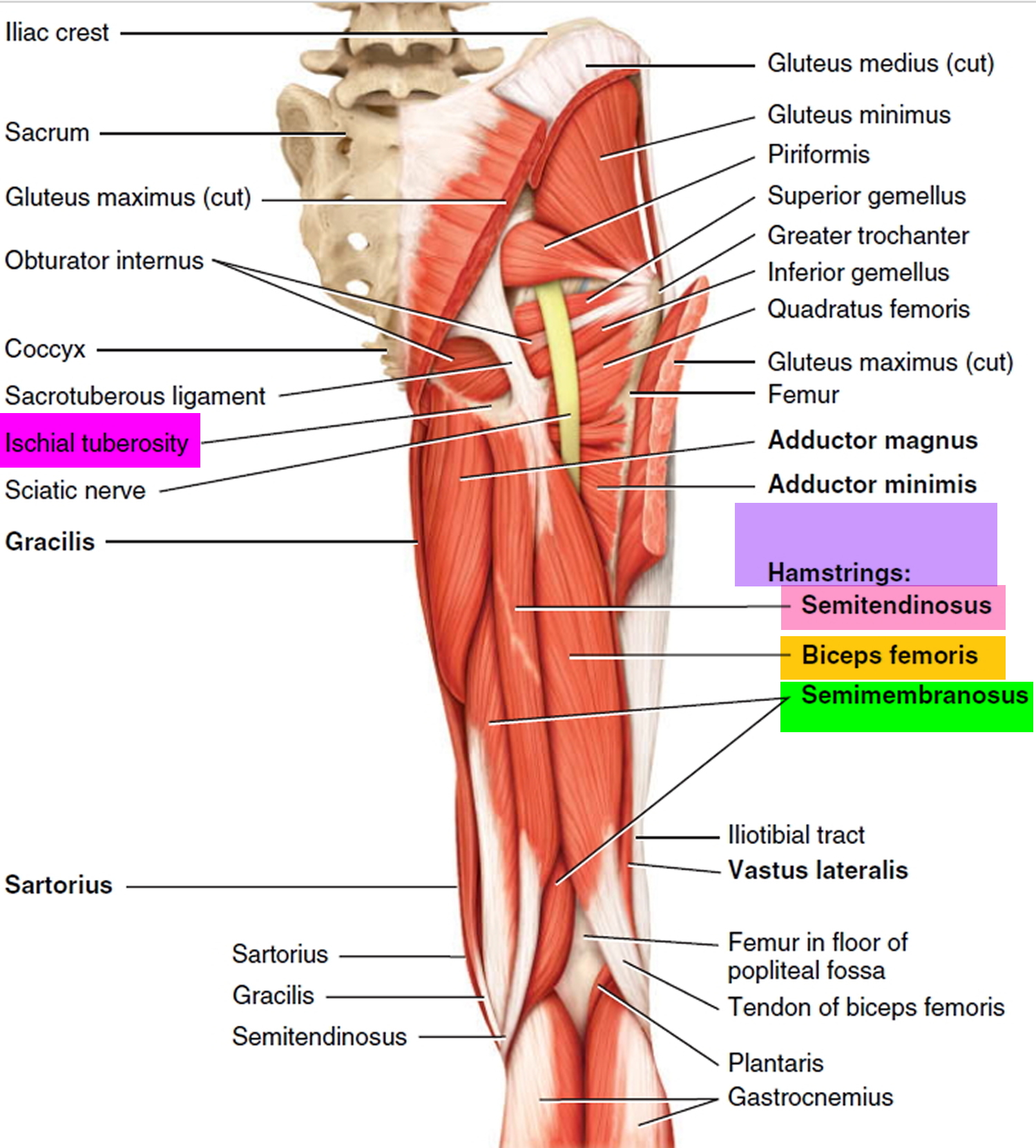
When any one of the three hamstring muscles is stretched beyond its limit hamstring strain can occur. The hamstrings are found medial to the gracilis and adductor magnus and lateral to the vastus lateralis.

Cruel masters of the middle ages severed the hamstrings of domestic slaves or prisoners in order to curtail escape.

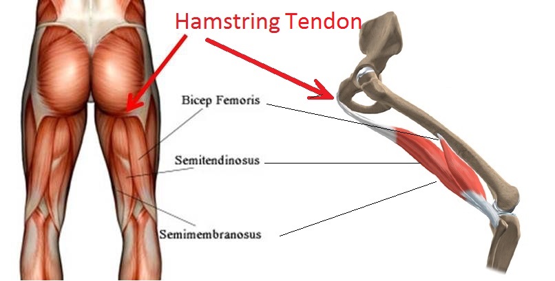
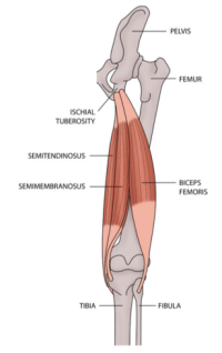
Anatomy hamstring. Learn how to begin treating a hamstring pull or tear and reduce your time on the sidelines. These muscles originate just underneath the gluteus maximus on the pelvic bone and attach on the tibia. Anatomy of the hamstring muscles.
Anatomy of an injury. Muscles should be inserted over the knee joint in the tibia or in the fibula. As group these muscles act to extend at the hip and flex at the knee.
The three hamstring muscles alone make up the classification of muscles known as the posterior compartment of the thigh. Muscles should originate from ischial tuberosity. Hamstring strains tend to be either the result of sudden stopping and starting during a sport sprinting for example or extreme stretching as might occur in gymnastics dance or yoga.
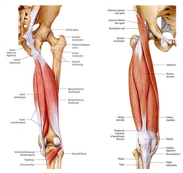
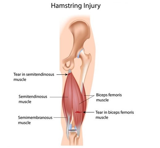
The muscles in the posterior compartment of the thigh are collectively known as the hamstrings. All three attach by long tendons crossing the back of the knee to the lower leg. There are two hamstrings on the medial inner side of the back of the thigh and one on the lateral outer side.
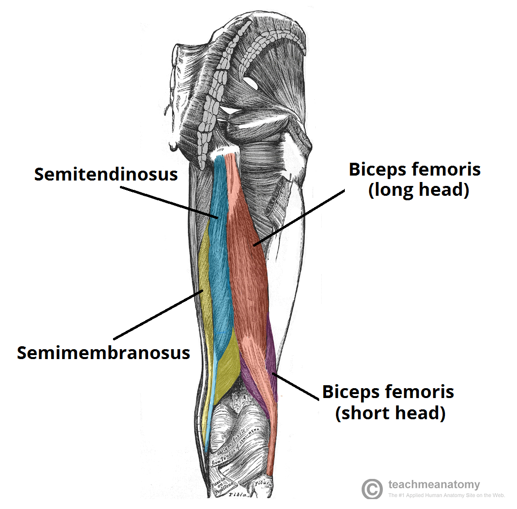
Muscles will be innervated by the tibial branch of the sciatic nerve. The hamstrings lie in the superficial muscle layer of the posterior thigh with the semitendinosus a and semimembranosus b on the medial side and the long head c and e and short head d of the biceps femoris on the lateral side of. Muscle will participate in flexion of the knee.
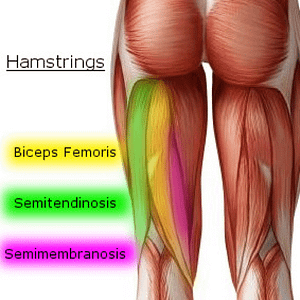
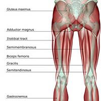
They consist of the biceps femoris semitendinosus and semimembranosus which form prominent tendons medially and laterally at the back of the knee. The biceps femoris semitendinosus and semimembranosus. The common criteria of any hamstring muscles are.
Recovery should begin the moment after an injury occurs. The hamstrings are comprised of three separate muscles. The hamstrings refer to 3 long posterior leg muscles the biceps femoris semitendinosus and semimembranosus.
Hamstring pulls and tears. Each originates attaches on the sitting bones of the pelvis and runs down the back of the thigh. These muscles start at the bottom of your pelvis extending down the back of your thigh and along either side of your knee to your lower leg bones.
Aspetar Sports Medicine Journal Review Of Hamstring Anatomy
 Basic Anatomy Of How To Stretch The Hamstrings The
 Hamstring Injuries Victorian Wolves Supporters Club
 Lower Anatomy Knee Hamstring Diagram Quizlet
 Knee Pain Caused By A Hamstring Injury Wayne Nj High

 Hamstring Tear Ortho Illinois
 Intramuscular Hamstring Tendon Injury Prognosis Surgical
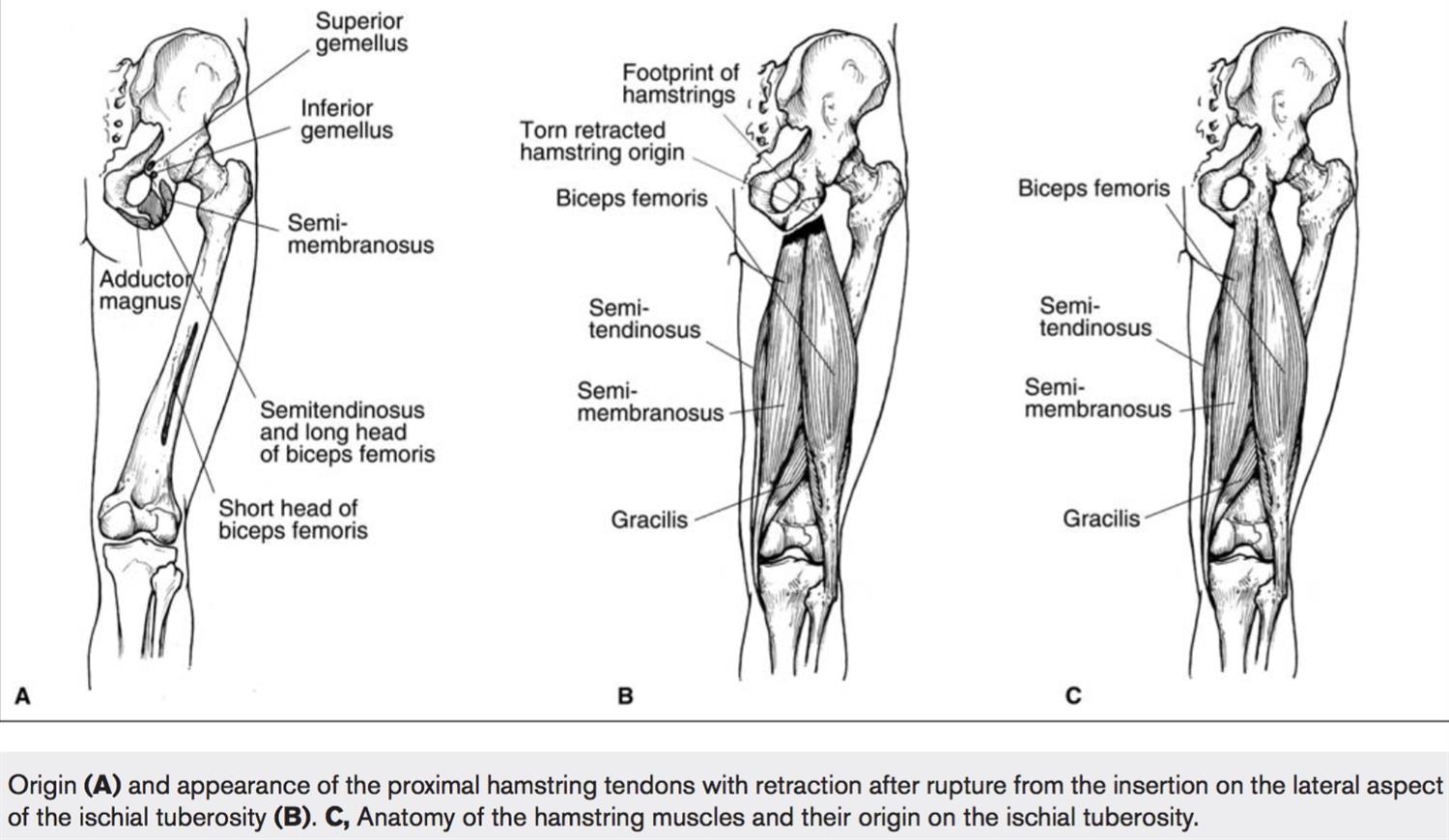
 Pdf Hamstring Injuries Anatomy Imaging And Intervention
 A Pain In The Rear High Hamstring Tendinitis Runner S World
 The Hamstrings Yoga Anatomy
 Hamstring Injury Local Physio
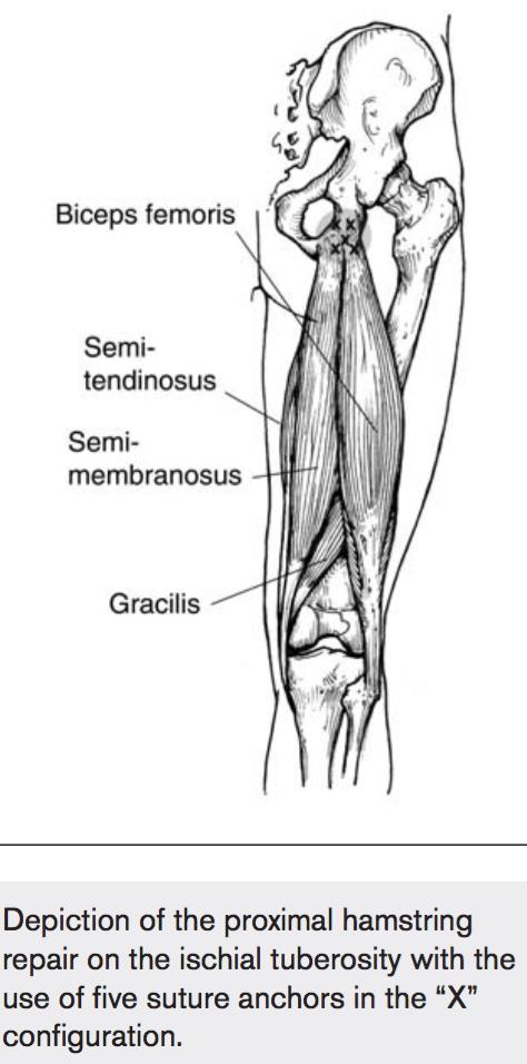
 Figure 1 From Rehabilitation And Prevention Of Proximal

 Illustration Picture Of Medical Anatomy Hamstring Muscle
 Hamstring Strain Exercises From A Pilates Perspective
 Hamstring Wikipedia
 Hamstring Injuries Knee Sports Orthobullets
 Hamstrings 201 The Advanced Course Fleet Feet Columbus
 Hamstring Muscles 1 Semitendinosus 2 Semimembranosus 3
:max_bytes(150000):strip_icc()/Depositphotos_19871399_original-56a05f523df78cafdaa14cd1.jpg) Hamstring Muscles
Hamstring Muscles
How Many Insertions Does The Hamstring Muscle Have Quora
 Hamstring Injuries Knee Sports Orthobullets
 Hamstring Injury Symptoms Recovery Treatment
 Muscles Of The Posterior Thigh Hamstrings Damage
 Hamstring Injury Causes Symptoms Recovery Time Treatment
Belum ada Komentar untuk "Anatomy Hamstring"
Posting Komentar