Foot Anatomy Of Bones
Foot and ankle anatomy is quite complex. The phalanges which are the bones in your toes.
 Anatomy Of The Bones Of The Foot The Bmj
Hind means posterior so it basically the backward part of the foot.
:watermark(/images/watermark_only.png,0,0,0):watermark(/images/logo_url.png,-10,-10,0):format(jpeg)/images/anatomy_term/talus/mh2AAjwGZlUw2Pt1lhUhQ_rvp4tyYMWn_Talus.png)
Foot anatomy of bones. Learn this topic now at kenhub. The hindfoot forms the heel and ankle. The talus which is the.
This is an article covering the muscle attachments blood supply innervation and ossification of the phalanges of the foot. The hindfoot consists of bone from the leg and the ankle joint. The foot is located after the long shin bones and it starts from the back of your ankle to your toes.
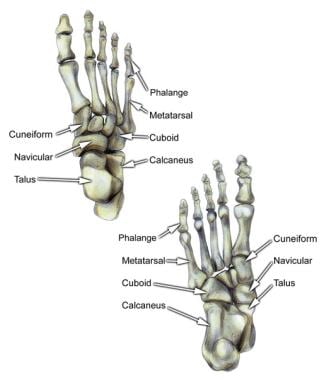
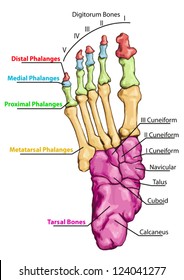
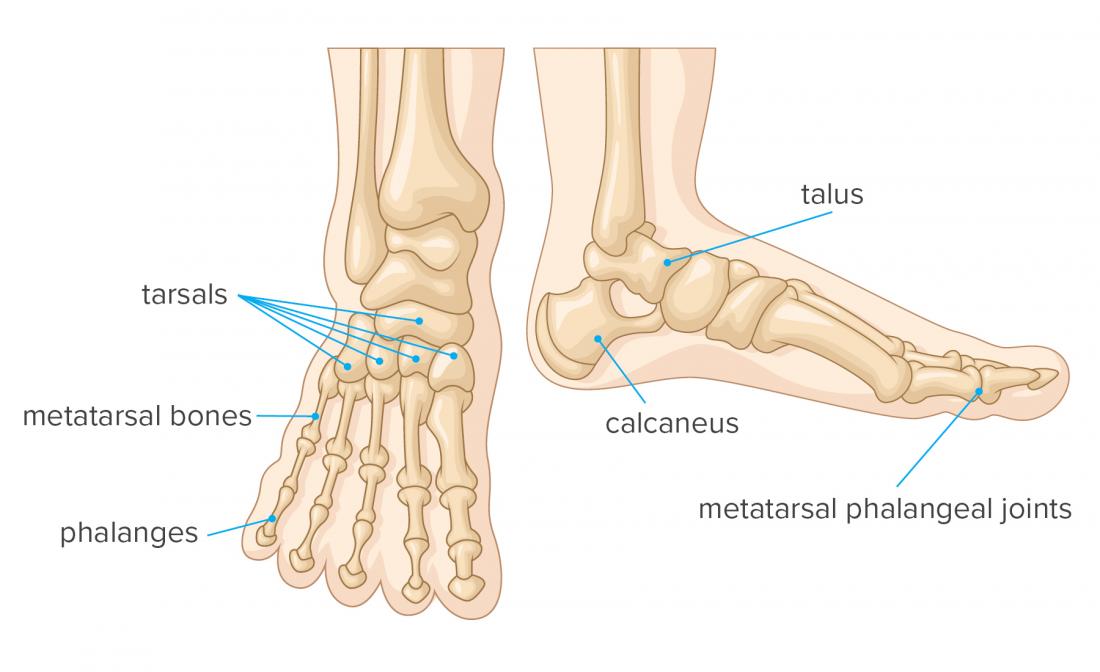
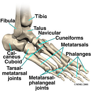
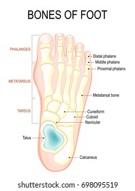
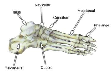
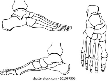
Anatomically the foot is divided into 3 sections. Parts of foot bones. The bones of the foot are organized into the tarsal bones metatarsal bones and phalanges.
This is an article covering the articular surfaces ligaments and muscles that produce movement at the joints of the feet. The different bones on each section of the foot. The foot is comprised of many bones joints tendons and ligaments including the plantar fascia and the achilles tendon.
Learn about the anatomy of the foot. The other bones of the foot that create the ankle and connecting bones include. The forefoot contains the five toes phalanges and the five longer bones metatarsals.
These all work together to bear weight allow movement and provide a stable base for us to stand and move on. The midfoot is a pyramid like collection of bones that form the arches of the feet. The feet are divided into three sections.
The foot begins at the lower end of the tibia and fibula the two bones of the lower leg. The parts of the foot bones. The hindfoot midfoot and the forefoot.
The calcaneus which is the bone in your heel. Bones and main ligaments of the foot. The cuneiform bones the navicularis and the cuboid all of which function to give your foot.
The talus bone supports the leg bones. The metatarsals which run through the flat part of your foot. At the base of those a grouping of bones form the tarsals which make up the ankle and upper portion of the foot.
The foot consists of thirty three bones twenty six joints and over a hundred muscles ligaments and tendons. The hindfoot is the posterior part of the foot. The seven tarsal bones are.
 Bones Of The Human Foot With The Name And Description Of All
 Foot Bone Anatomy Overview Tarsal Bones Gross Anatomy
 Foot Vertebrate Anatomy Britannica
 Foot Bones Photos 40 827 Foot Stock Image Results
 Skeletal System Anatomy Bones Of The Ankle Foot And Toes
Anatomy Physiology Illustration
 Bones Of The Foot Quiz Anatomy
 Bones Stock Illustrations 42 153 Bones Stock Illustrations
 Foot Bones Photos 40 827 Foot Stock Image Results
:watermark(/images/watermark_only.png,0,0,0):watermark(/images/logo_url.png,-10,-10,0):format(jpeg)/images/anatomy_term/talus/mh2AAjwGZlUw2Pt1lhUhQ_rvp4tyYMWn_Talus.png) Ankle And Foot Anatomy Bones Joints Muscles Kenhub
Ankle And Foot Anatomy Bones Joints Muscles Kenhub
 Foot Bones Anatomy Conditions And More
 Houseables Disarticulated Human Skeleton Full Anatomical Model Life Sized 62 Height Plastic With Poster Skull Bones Articulated Hand Foot
 Foot And Ankle Orthopedics Seaview Orthopaedic Medical
 Foot Bones Photos 40 827 Foot Stock Image Results
 Human Skeleton Body Parts Skull Bones Hands Foot
Bones Of The Leg And The Foot Skeleton Of The Hindlimb
 Foot Bone Anatomy Overview Tarsal Bones Gross Anatomy
 Foot Bones Photos 40 827 Foot Stock Image Results
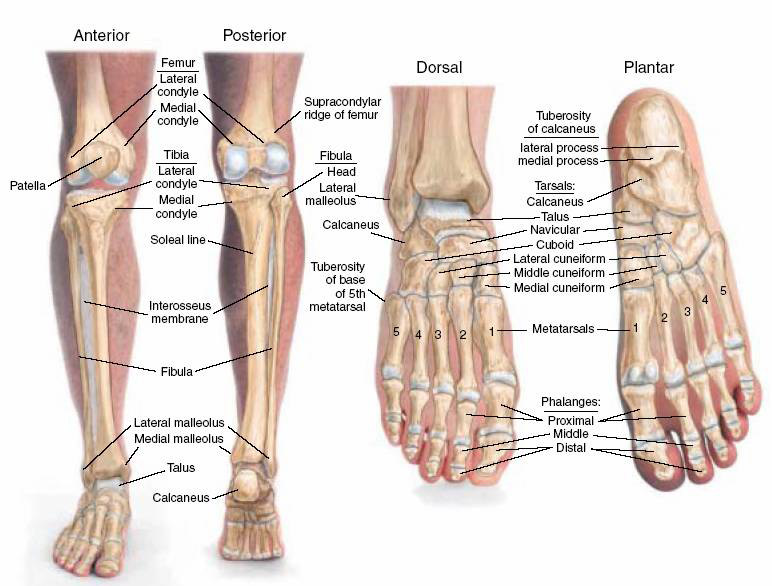 The Bones Of The Leg And Foot Anatomy Medicine Com
 Axis Scientific Complete Disarticulated Human Skeleton Bundle Includes 3 Part Human Skull Life Size Bones Articulated Hand And Foot Anatomy


Belum ada Komentar untuk "Foot Anatomy Of Bones"
Posting Komentar