Human Sinuses Anatomy
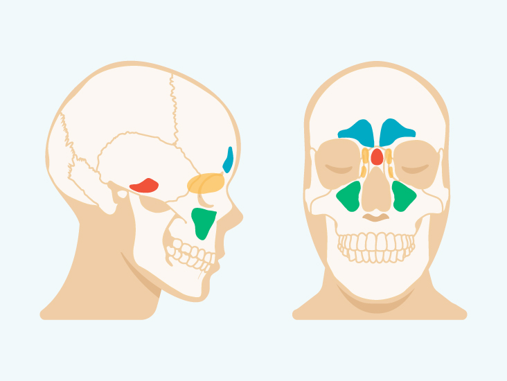
The maxillary and ethmoid sinuses are present at birth starting to form around the 3rd or 4th month of gestational development 10. The ethmoid sinuses lie under the inside corners of the eyes.
 Cross Section At The Level Of The Frontal Sinus Preview Human Anatomy Kenhub
The sinuses are a connected system of hollow cavities in the skull.

Human sinuses anatomy. As in the nasal passage the sinuses are lined with mucous membranes. The word sinusitis is used to indicate that one or more of the membrane linings found in the sinus cavities has become inflamed or infected. The maxillary sinuses are located on.
Others are much smaller. Anatomy of the paranasal sinuses development. The sphenoid sinuses are located behind the ethmoid sinuses.
The largest sinus cavities are about an inch across. It is however distinct from a fistula which is a tract connecting two epithelial surfaces. The paranasal sinuses are connected to the nasal cavity through small orifices called ostia.
The sinuses are hollow spaces in the skull and the face bones around your nose. This sinus is located inside the face around the area of the cheeks. They further develop over the first few years of life 11.
There are four pairs of sinuses named for the bones that theyre located in. It is present at birth and continues to grow. The frontal sinuses lie behind the forehead above the eyes.
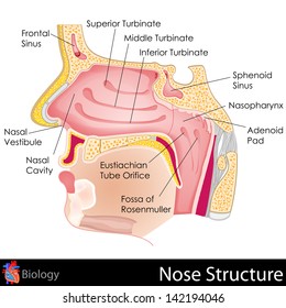
Sphenoethmoidal recess is a small area above the superior concha that receives the opening of the sphenoid sinus. The sinuses are lined with mucus producing membranes that help guard against pathogens debris and pollutants. This sinus is located inside the face around the area of the bridge of the nose.
The maxillary sinuses are behind the cheeks. There are four different types of sinuses. In anatomy the term is used in various contexts.
Rudimentary sphenoid sinuses are there at birth forming pneumatizing completely by the age of 5 years 6. Your cheekbones hold your maxillary sinuses the.
 Inferior Sagittal Sinus Human Anatomy Kenhub
 Big Nasal Cavity Human Anatomy Sinus With Face Model Buy Human Anatomy Model Nasal Cavity Model Human Nasal Model Product On Alibaba Com
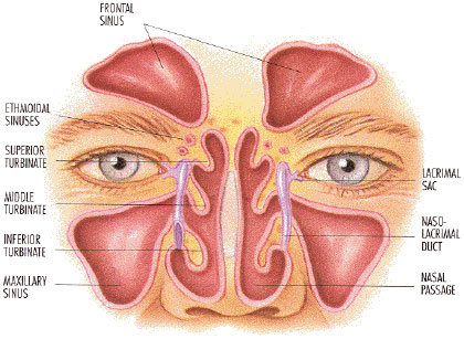
 Sinuses Of Nose Human Anatomy Sinus Diagram Anatomy Of The
 The Paranasal Sinuses Structure Function Teachmeanatomy
 Equine Sinus Disease A Hidden Danger Expert How To For
 Sinus Cavities In The Head Anatomy Diagram Pictures
 Figure Anatomy Of The Paranasal Sinuses Spaces Between The
 Anatomy Of Sinuses Brain Sinus Anatomy Human Anatomy Diagram
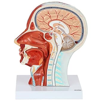 Axis Scientific Human Head Model Anatomy Model Features Half Head Muscular Anatomy Veins Arteries Exposed Sinuses Brain And Spinal Anatomy
 Sinus Cavities In The Head Anatomy Diagram Pictures
![]() Dural Venous Sinuses Anatomy Kenhub
Dural Venous Sinuses Anatomy Kenhub
 Sinuses Sinusitis Rhinosinusitis Defined Aaaai
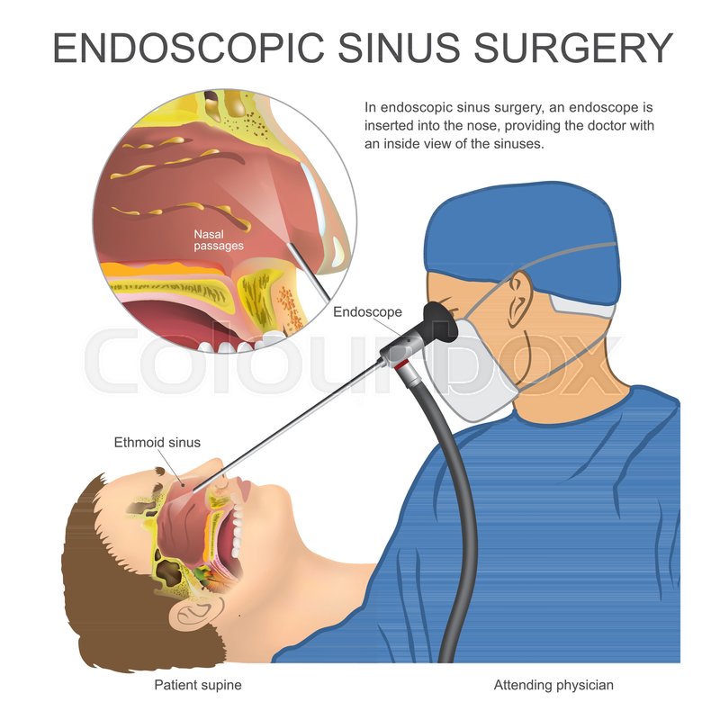 In Endoscopic Sinus Surgery An Stock Vector Colourbox
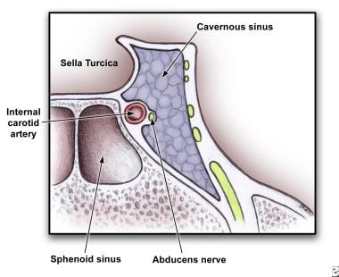 What Is The Anatomy Of The Cavernous Sinuses
 World S Best Paranasal Sinus Stock Illustrations Getty Images
 Amazon Com Emvency Wall Tapestry Sinuses Of Nose Human
 Nose And Sinuses Anatomical Chart Laminated Card Science
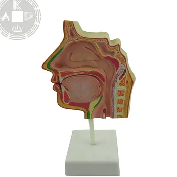
Eisco Labs Model Human Nose And Sinus Longitudinal Section
 Medical Infographic Of Sinus And Human Nasal Anatomy
 Nose Anatomy Images Stock Photos Vectors Shutterstock
 Sinus Model Human Body Anatomy Replica Of Normal Nose
 Vector Illustration Nose Sinus Anatomy Human Respiratory System
 Nose And Sinus Models
 Amazon Com Human Brain Head Model Anatomy Model Features
 Us 111 18 49 Off Human Head Anatomical Model Skull Anatomy Sagittal Sinus Oral Nasopharyngeal Medical Teaching Model In Medical Science From Office
 Acute Sinusitis A Cost Effective Approach To Diagnosis And
 Understanding Sinusitis Chart 22x28 Clinicalposters
Nasal Sinus Program Davis Ear Nose Throat Sleep Center


Belum ada Komentar untuk "Human Sinuses Anatomy"
Posting Komentar