Shoulder Arthroscopy Anatomy
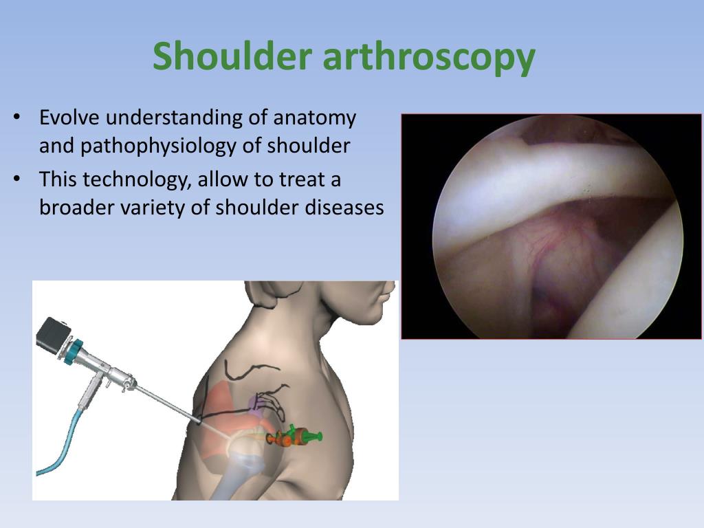
The anatomy of the shoulder. Shoulder arthroscopy is an option for a variety of shoulder conditions.
 Ppt Shoulder Arthroscopy Powerpoint Presentation Free
Center for sports medicine and orthopaedics chattanooga tn shoulder arthroscopy normal anatomy review.
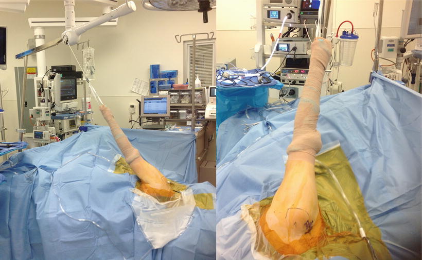
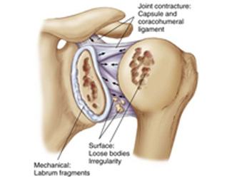
Shoulder arthroscopy anatomy. The glenohumeral joint is where the ball humeral head and the socket the glenoid meet. The surgical arm is then placed in a sling with longitudinal traction. Your upper arm bone humerus your shoulder blade scapula and your collarbone clavicle.
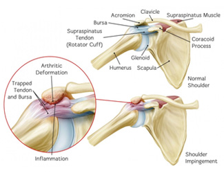
The acromioclavicular joint is where the acromion part of the shoulder blade scapula and the collar bone clavicle meet. It is made up of three bones. The introduction of shoulder arthroscopy has meant that a thorough knowledge.
The head should be secured in a neutral position and the contralateral elbow and knees in flexion. Commonly a pulley system is used where the surgical arm is suspended in the air by a sling connected by a rope. Shoulder arthroscopy anatomy your shoulder is a complex joint that is capable of more motion than any other joint in your body.
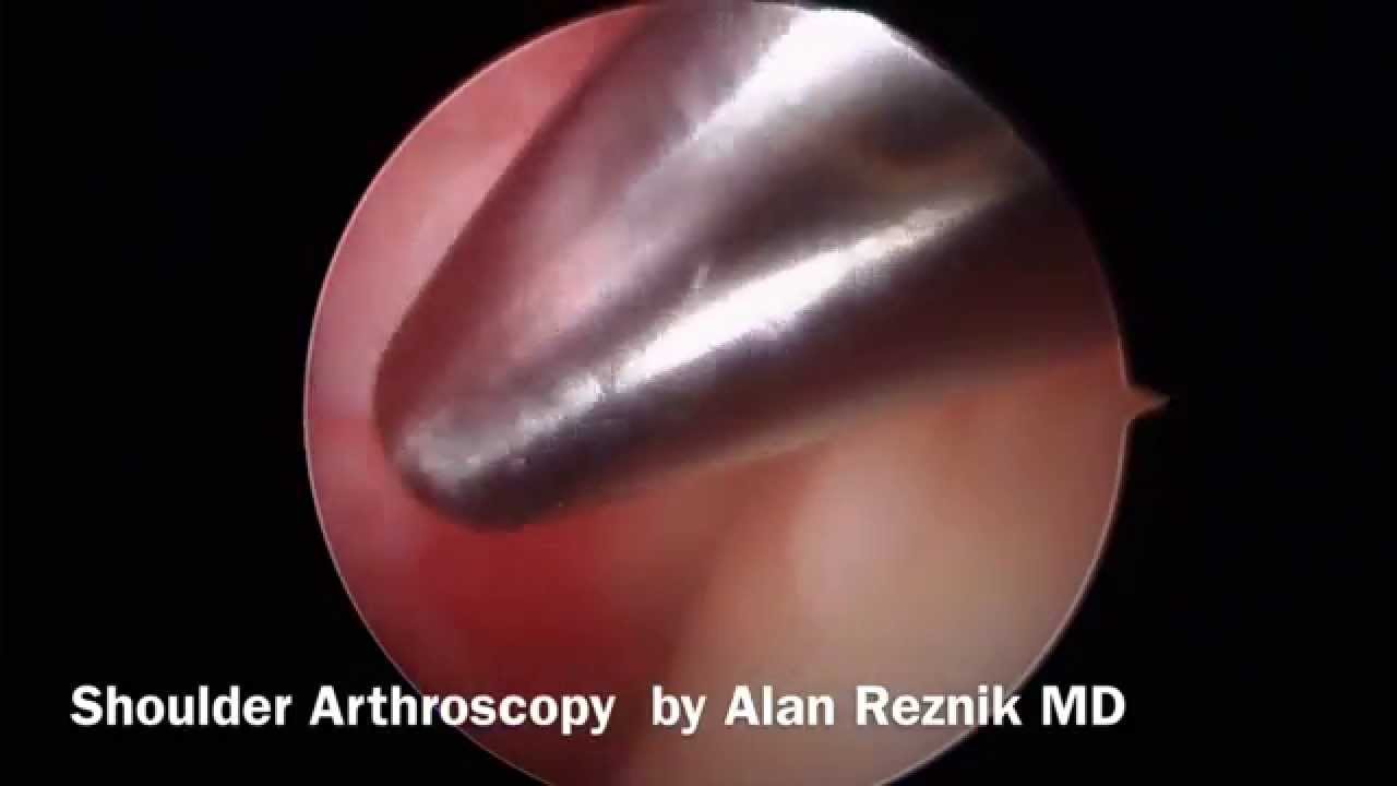
It is the only imaging modality that allows the internal anatomy of the shoulder to be directly visualised in the living patient whereas other imaging techniques allow shoulder anatomy to be viewed from an outside in perspective. The acromioclavicular joint where the acromion of the shoulder blade and the collarbone clavicle meet and the glenohumeral joint where the head of the humerus the upper bone in the arm meets the glenoid the cup like portion of the scapula. Diagnostic shoulder arthroscopy and arthroscopic anatomy.
The shoulder is made up of two joints the acromioclavicular joint and the glenohumeral joint. Options in shoulder arthroscopy. Shoulder arthroscopy can address problems with any part of the joint anatomy to provide diagnosis and sometimes treatment without more invasive surgery.
Anatomy of the shoulder joint while many people think of the shoulder as a single joint it is actually made up of two joints.
 Shoulder Arthroscopy Anatomy And Variants Part 2
 Shoulder Arthroscopy Anatomy And Variants Part 2
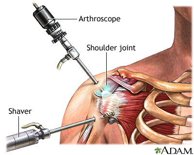
 Shoulder Arthroscopy
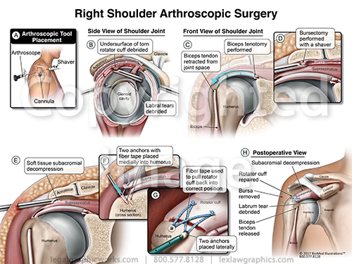 Right Shoulder Arthroscopic Surgery
 Diagnostic Shoulder Arthroscopy And Arthroscopic Anatomy
 Update Your Understanding Of Shoulder Arthroscopy Codes
 What Is A Rotator Cuff Repair Shoulder Arthroscopy Colorado Shoulder Surgeon
 An Introduction To Shoulder Arthroscopy
 Arthroscopic Shoulder Surgery Anatomy Basic To Advanced
 Shoulder Arthroscopy Conditions Treated
 Intro To Shoulder Arthroscopy Rotator Cuff Tear Anatomy
 Shoulder Arthroscopy Arthroscopic Subacromial Bursectomy
 Shoulder Arthroscopy
 Shoulder Joint Capsule Tissue Picture Shoulder Joint
 Mini Open Rotator Cuff Repair The Hughston Clinic

 Figure 3 From Shoulder Arthroscopy Positioning Lateral
 Shoulder Arthroscopy

 Arthroscopic Anatomy Of Shoulder Springerlink
 Glenohumeral Arthroscopy Beach Chair Position Left
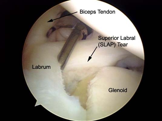
 Shoulder Arthroscopy Normal Anatomy
 Dr Prohaska Shoulder Arthroscopy Advanced Orthopedic

Shoulder Arthroscopy Information Mount Sinai New York
Belum ada Komentar untuk "Shoulder Arthroscopy Anatomy"
Posting Komentar