Anatomy Of The Heart And Blood Flow
Each valve has a set of flaps called leaflets or cusps. This is the process called oxygenation.
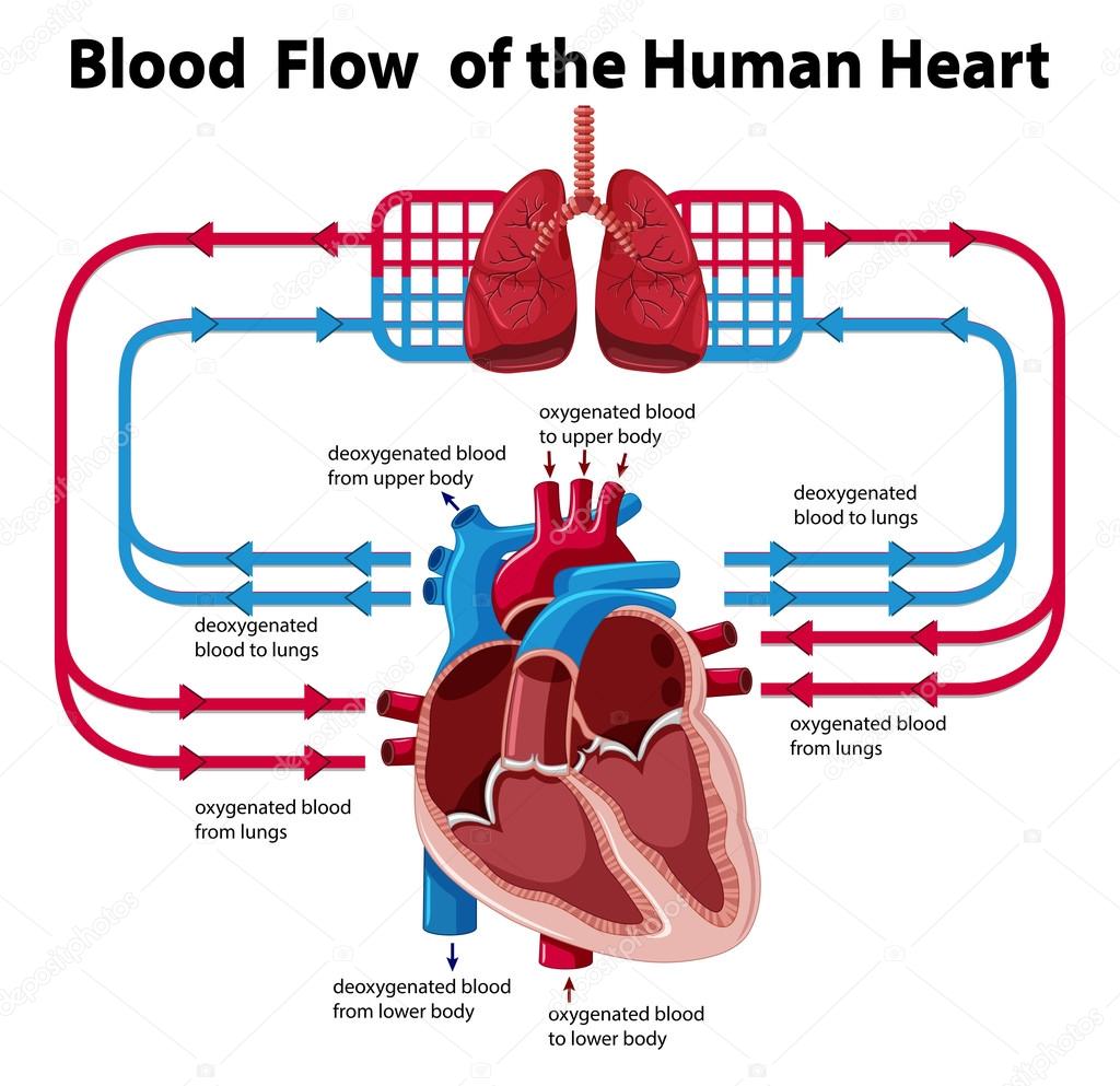 Clipart Heart Anatomy Chart Showing Blood Flow Of Human
The aortic and pulmonic valves lie between the ventricles and the major blood vessels leaving the heart.

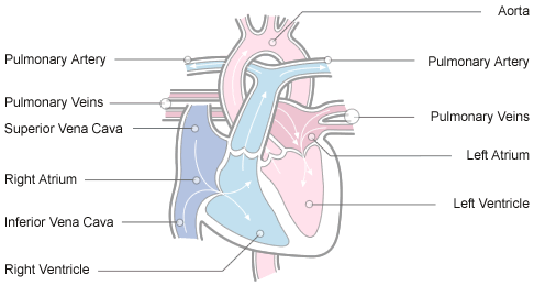
Anatomy of the heart and blood flow. Blood enters the heart through two large veins the inferior and superior vena cava emptying oxygen poor blood from the body into the right atrium of the heart. However the blood flow through the heart is a little different. Heart valves are controlled by pressure changes within each chamber and contraction and relaxation are controlled by the hearts conduction system.
The heart is the fluid pump of the cardiovascular system. The flow of blood through the heart is controlled by the opening and closing of valves and the contraction and relaxation of the myocardium. They prevent blood from flowing in the wrong direction.
There are six structures on each of these sides that are used during blood. Blood now moves from the right ventricle through the pulmonary semilunar valve which is the valve that separates the right ventricle and the lungs. The size of your heart can vary depending on your age size and the condition of your heart.
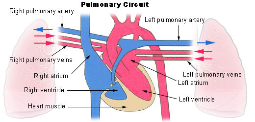
As we previously learned this is called the pulmonary circuit. The heart valves work the same way as one way valves in the plumbing of your home. In the lungs the blood picks up oxygen and drops off carbon dioxide before returning to the heart.
Anatomy of the heart. Blood is conducted through blood vessels arteries and veins. Its muscular walls beat or contract pumping blood to all parts of your body.
It pumps blood and nutrients to all of the organs of the body. Right side of the heart. Blood is prevented from clotting in the blood vessels by their smoothness and the finely tuned balance of clotting factors.
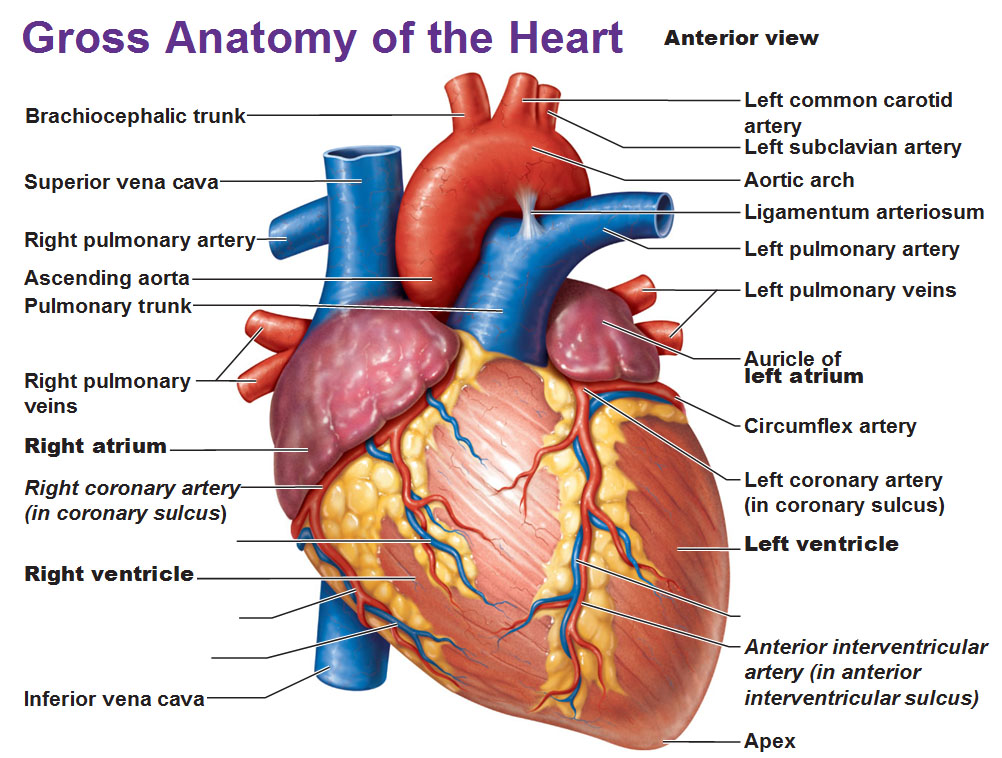
The left side and the right side. The valves are also shown including the tricuspid bicuspid mitral aortic and. Your heart is located under your ribcage in the center of your chest between your right and left lungs.
And it pumps blood to the lungs where it receives oxygen after the body has used it up. The chambers of the heart are shown including the right atrium left atrium right ventricle and left ventricle. When learning the blood flow of the heart it helps to divide it up into two sections.
Its included in this module as one of the steps in the heartbeat cycle.
 Three Chambered Heart Definition Anatomy
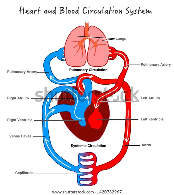 Blood Circulation System Stylized Heart Anatomy Stock Vector
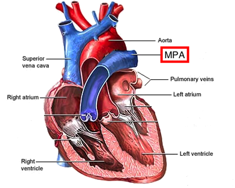 Blood Flow In The Arteries Practice Khan Academy
About Your Heart How Does Blood Flow Through The Heart
The Reptipage Crocodylian Bodyplans
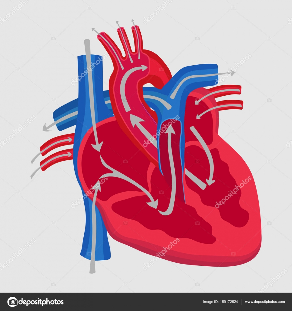 The Human Heart The Study Of Anatomy The Path Of Blood
 Blood Pathway Of Heart Anatomy Cardiac Nursing Nursing
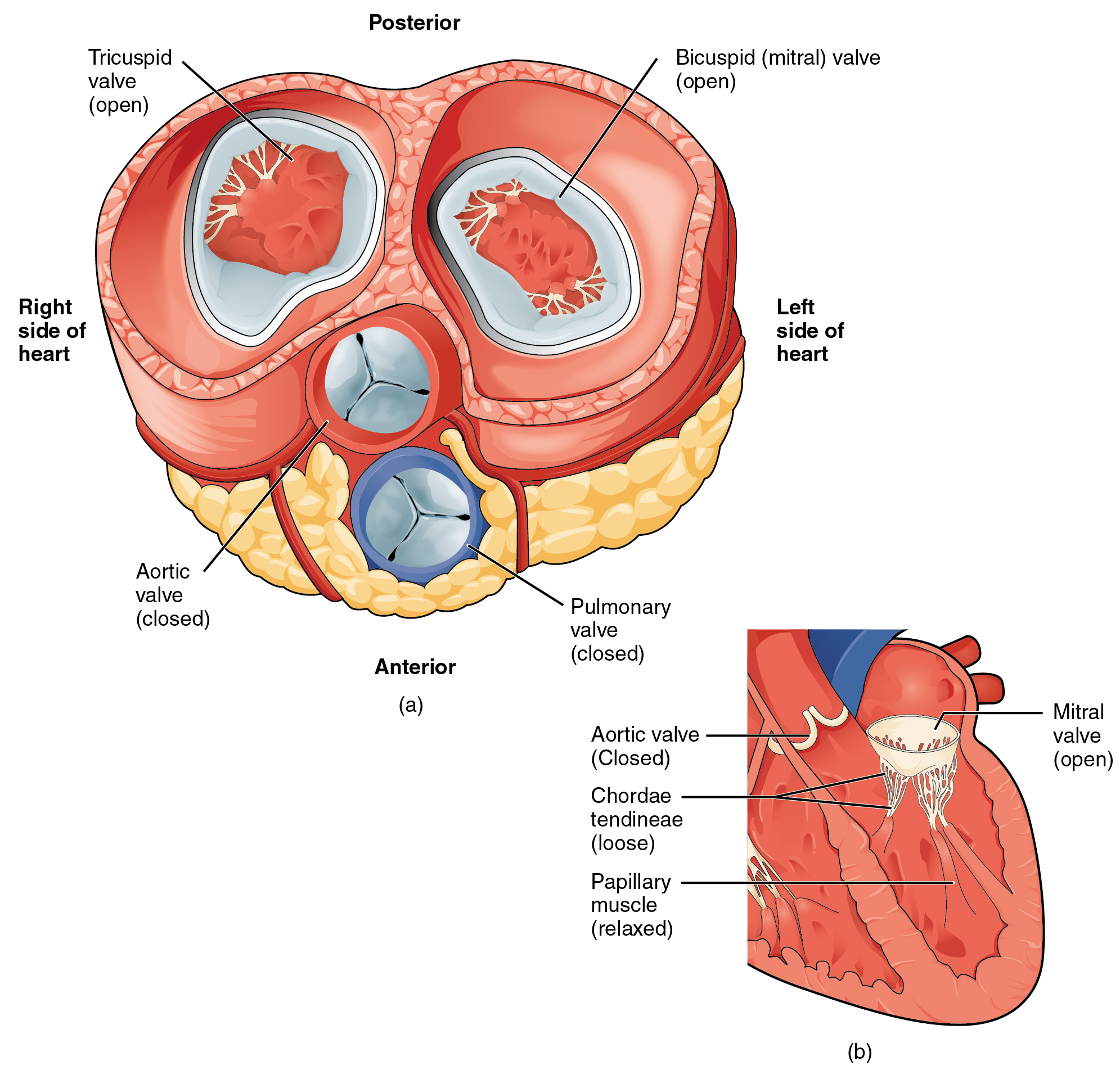 19 1 Heart Anatomy Anatomy And Physiology
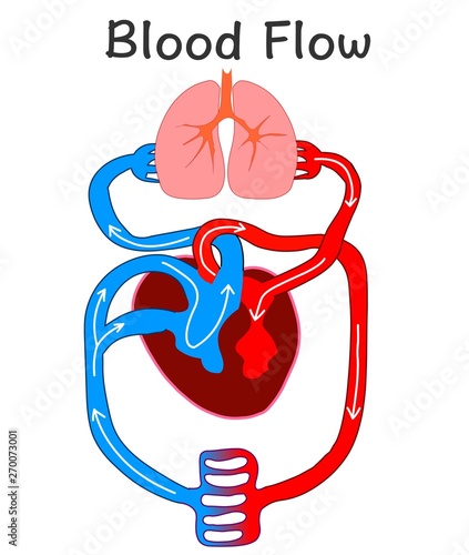 Blood Flow Heart Anatomy Formation Human Circulation

 Pin By Espinoza Hosa On Essentials Of Healthcare Heart
 Amazon Com Blood Flow Through The Heart Paper Print Wall
 Heart Anatomy Blood Flow Booklet
 Blood Flow Of The Heart By Megan Wiggins The Heart Anatomy
 Biology Blood Circulation Structure Of Human Heart
 Circulatory System Blood Flow Pathway Through The Heart
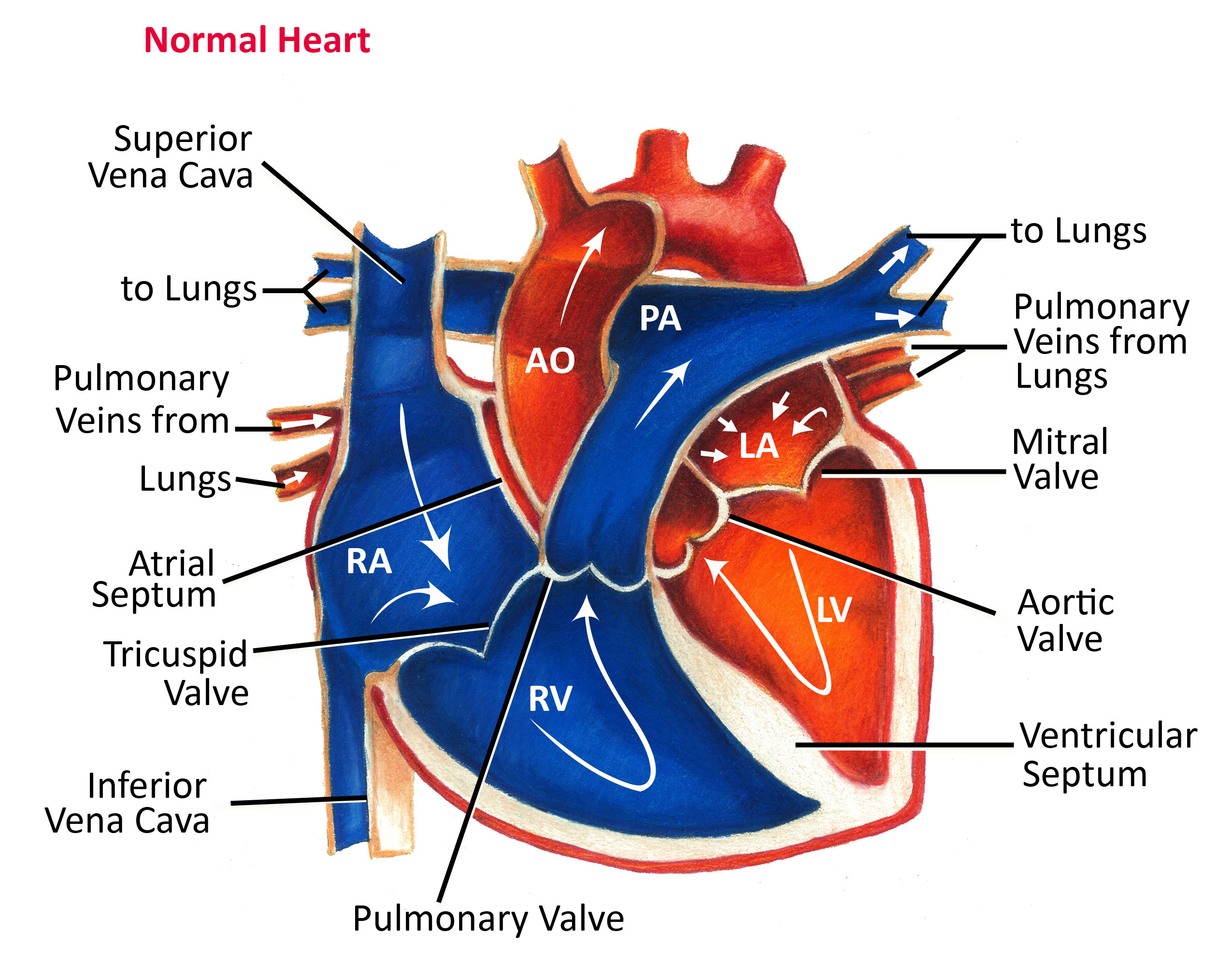 Normal Heart Anatomy And Blood Flow Pediatric Heart
 Cardiology Statistic Cardiovascular Organ Anatomy Blood Flow
 Pdf Augmented Reality To Teach Human Heart Anatomy And
 Seer Training Circulatory Pathways
 Cv Physiology Coronary Anatomy And Blood Flow

 The Circulatory System Junqueira S Basic Histology Text
 Blood Flow In Human Heart Realistic Vector Scheme
 Chapter 20 Module 1 Anatomy And Blood Flow Of The Heart
 Anatomy And Physiology Of The Heart Normal Function Of The
 Coronary Vessels Anatomical Health Care Vector Illustration
 Blood Flow Through The Body Boundless Anatomy And Physiology

Pediatricepsociety Anatomy Of A Healthy Heart


Belum ada Komentar untuk "Anatomy Of The Heart And Blood Flow"
Posting Komentar