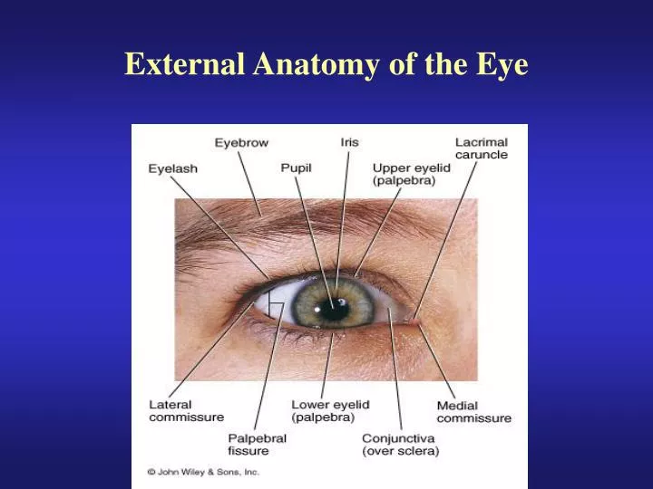
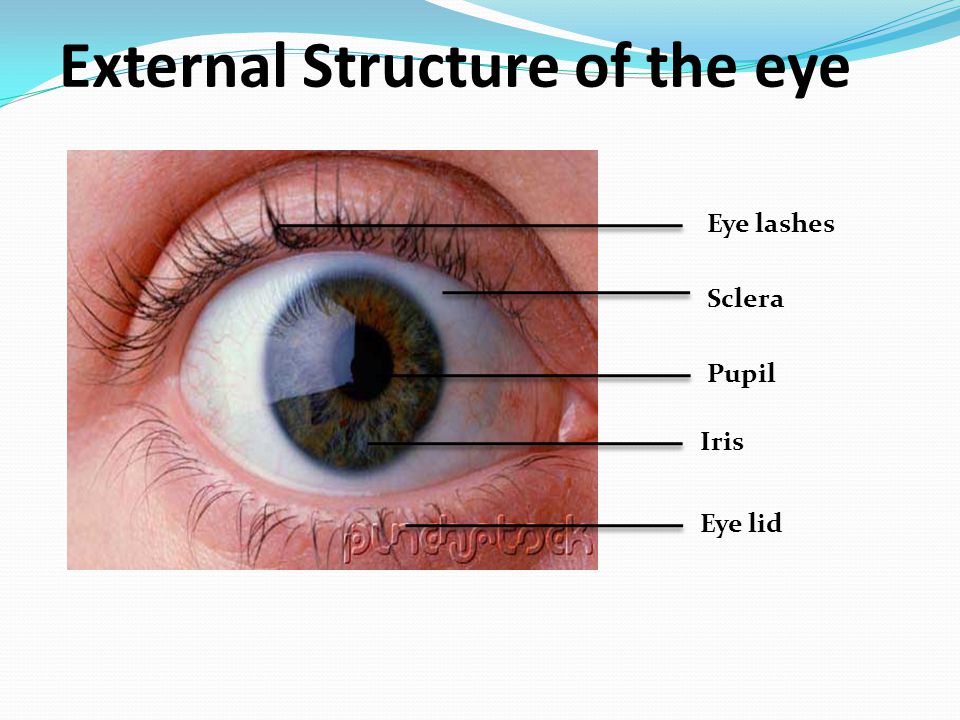
External Eye Anatomy
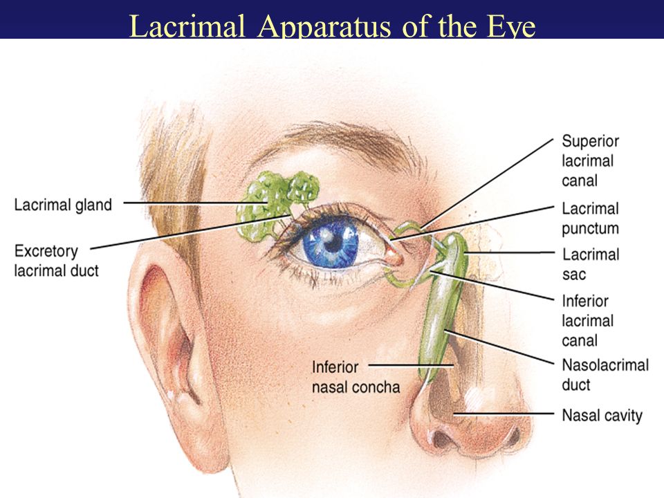
Lacrimal system tear drainage system the lacrimal system is crucial for tear production and management which includes distribution of tears and draining excess tears. Extraocular muscles help move the eye in different directions.

Low to high sort by price.

External eye anatomy. Modified sweat glands between lashes. Six muscles attach to the outer surface of the eye and produce mucous membrane that lines the eyelids and outer surface of th two movableshades that further protect the eye from injury st modified sebaceous glands lubricates eye. Malfunction in any part of the system can cause serious complications.
It is the most visible part of the eye. Learn vocabulary terms and more with flashcards games and other study tools. As light passes through the eye the iris changes shape by expanding and letting more light through or constricting and letting less light through to change pupil size.
The iris is part of the uveal tractthe middle layer of the wall of the eye. Home eye anatomy illustrations external eye anatomy showing 112 of 15 results default sorting sort by popularity sort by average rating sort by newness sort by price. The eye sits in a protective bony socket called the orbit.
Tap on the image or pinch out and pinch in to resize the image. These muscles move the eye up and down and side to side and rotate the eye. The outer fibrous or sclera 2.
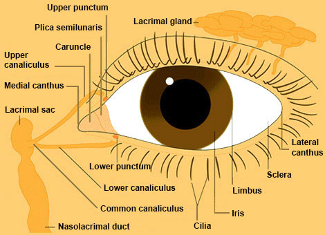
Nerve signals that contain visual information are transmitted through the optic nerve to the brain. Much less important than the lower punctum. This is a strong layer of tissue that covers nearly the entire surface of the eyeball.
It lies in front of the crystalline lens and separates the anterior chamber from the posterior chamber. The human eye ball is spherical in structure and is about 24 mm in a diameter. Six extraocular muscles in the orbit are attached to the eye.
The lens then changes shape to allow the accurate focusing of light on the retina. Oval opening in the upper lid margin where tears enter to flow to the lacrimal sac. The structure of the human eye is made of three layers.
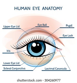
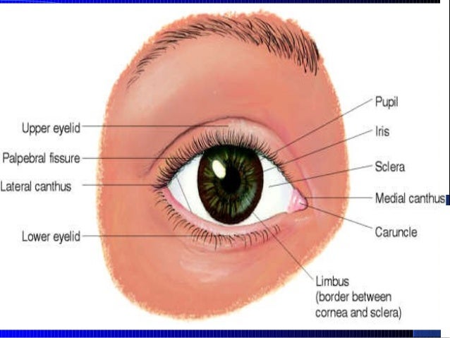
Anatomy of the eye. The extraocular muscles are attached to the white part of the eye called the sclera. The iris is the colored part of the eye that controls the amount of light that enters into the eye.
The eye is surrounded by the orbital bones and is cushioned by pads of fat within the orbital socket. Click on a label to display the definition. The cornea allows light to enter the eye.
Start studying external eye anatomy. Human eye parts 1.
 External Anatomy Of The Eye Ppt Video Online Download
 Human Eye Anatomy Structure Of The Eye


 Imagenes Fotos De Stock Y Vectores Sobre Eye Anatomy
 Eye External And Internal View
 Anatomy Of The Human Eye In Front External View Schematic Diagram
Parts Of The Eye American Academy Of Ophthalmology
 Stock Eye Anatomy Images From Jirehdesign Com Eye Illustrations
 Ppt External Anatomy Of The Eye Powerpoint Presentation
 Identify External Eye Anatomy Diagram Quizlet
 Cataract Surgery Symptoms Treatment Causes
 External Anatomy Human Eye Wall Mural Wallmonkeys Com

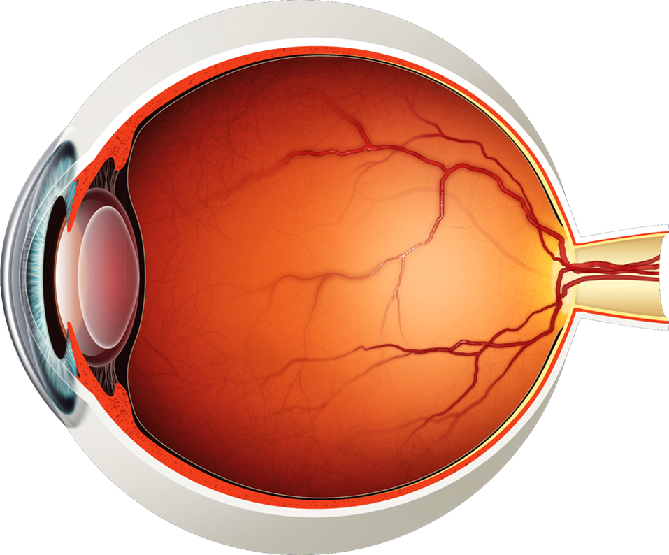 Diagram Of The Eye Lions Eye Institute
 Eye Anatomy Paediatric Academy
 Anatomy Of The Human Eye
 Aurolab Anatomy
 Styes Eye Infection Internal And External Hordeolum
 Eyes
Eyes
 Normal Eye Ultrasound How To
Parts Of The Eye American Academy Of Ophthalmology
 The External Structure Of The Eye Vector Illustration
Lateral View Of External Eye Anatomy Diagram Quizlet
 The External Structure Of The Eye
 The Eye Structure Function Ppt Video Online Download
 Identify External Eye Anatomy 2 Diagram Quizlet
Belum ada Komentar untuk "External Eye Anatomy"
Posting Komentar