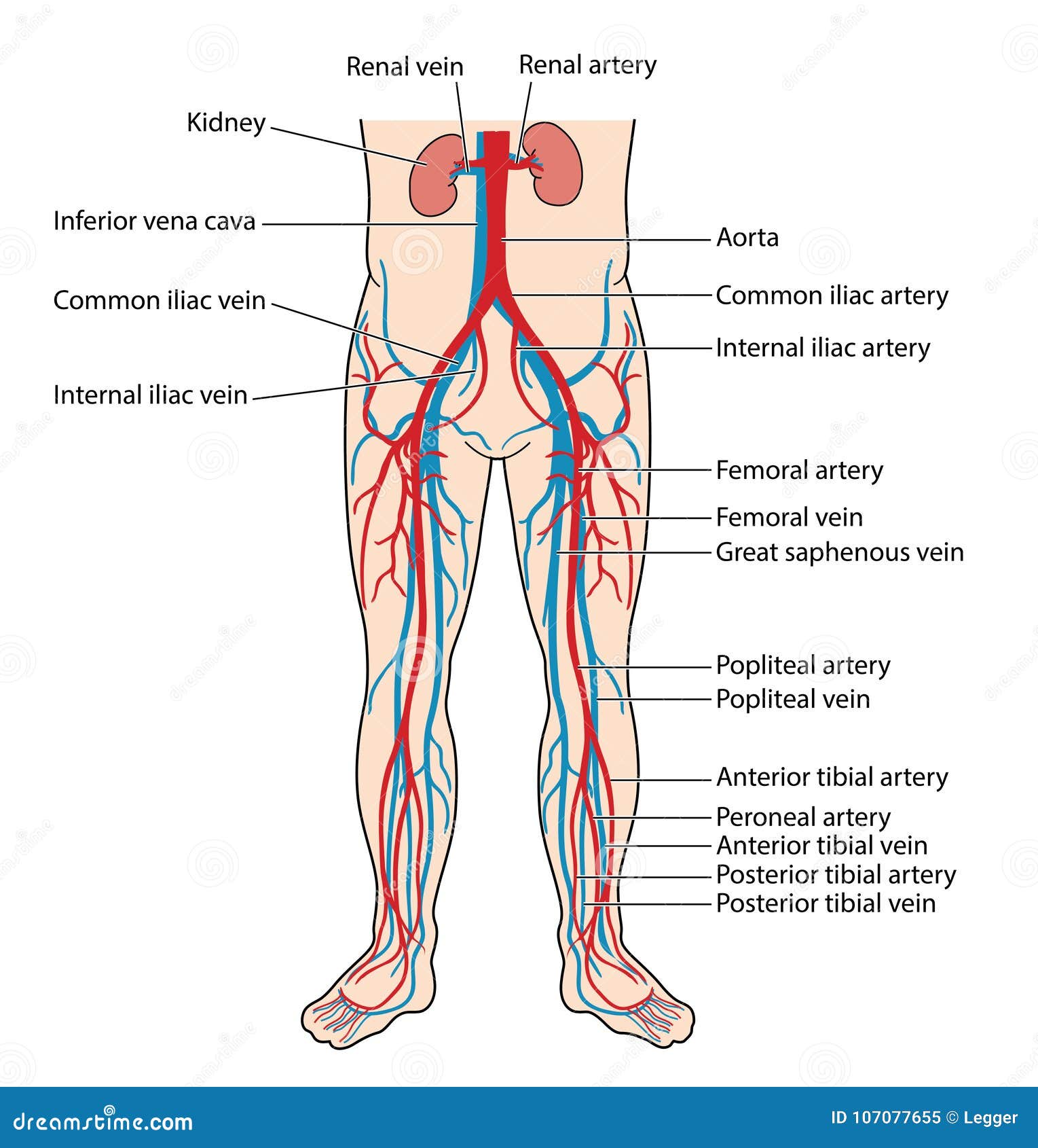
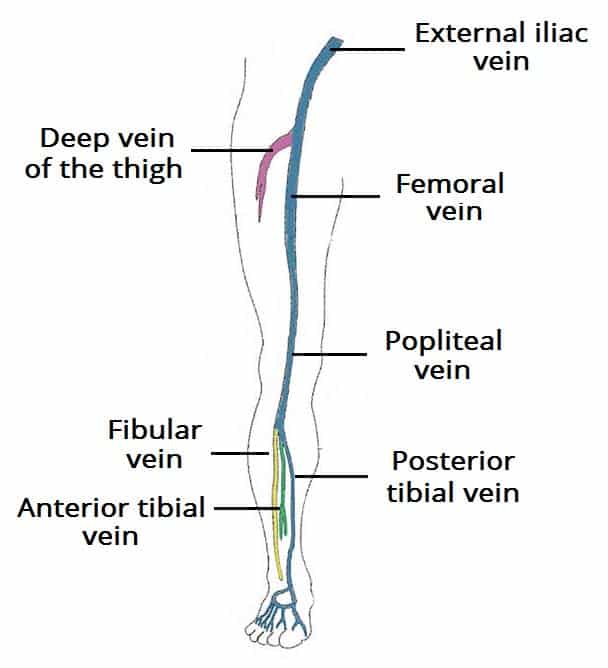
Leg Vein Anatomy
Place the probe transversely at the knee crease in the popliteal fossa. There are six of such tributaries.
 Assessment And Management Of Patients With Varicose Veins
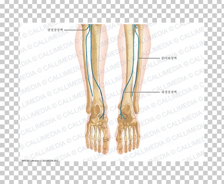
Deep veins of the foot form two divisions.
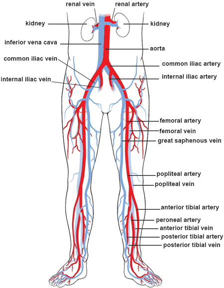
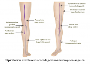
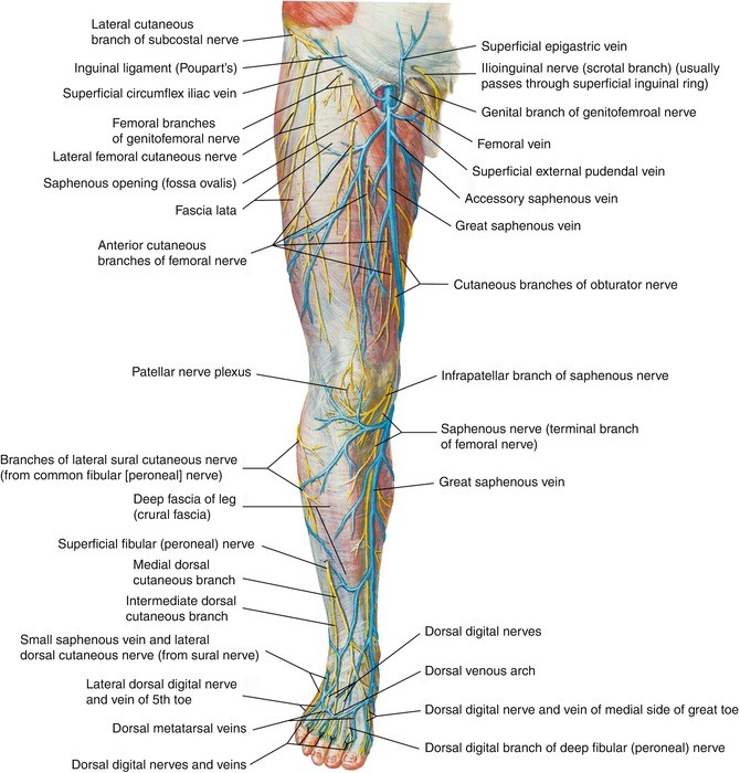
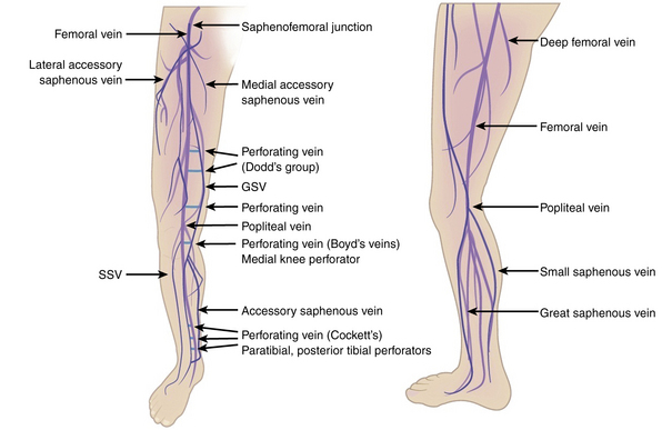
Leg vein anatomy. Seat the patient on the side of the bed to help dilate the veins for easier visualisation. It ascends up the medial side of the leg passing anteriorly to the medial malleolus at the ankle and posteriorly to the medial condyle at the knee. Posteromedial vein of the thigh accessory vein of the thigh drains the superficial aspect.
Anterior tibial vein which receives blood from the dorsal venous arch. Anterior femoral cutaneous vein a continuation of anterior veins in the distal thigh. In the context of diagnosing a deep vein thrombosis the posterior tibial and peroneal veins are the most frequently affected.
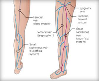
As the vein moves up the leg it receives tributaries from other small superficial veins. The great saphenous vein is formed by the dorsal venous arch of the foot and the dorsal vein of the great toe. Posterior tibial vein and fibular vein also known as the peroneal vein which form from the medial and lateral plantar veins.
Continue to follow the vein sequentially compressing down to the distal thigh. Superficial epigastric vein drains the inferior abdominal wall and opens. However their junctions are not paired and their locations are variable.
The plantar and the dorsal veins. The deep veins of the leg accompany the arteries of the same name and are generally paired in the calf. There are three main deep veins in the lower leg.
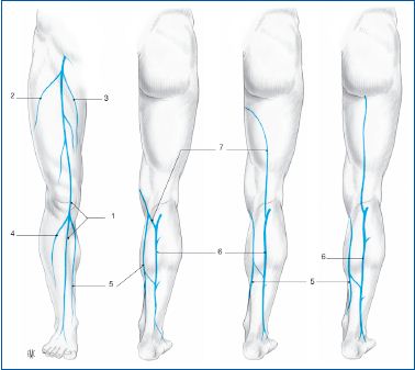
The lateral venous system is drained through multiple small tributaries into the gsv and ssv. Document the normal anatomy and any pathology found including doppler images demonstrating flow. The sural nerve courses along the ssv in the distal calf.
Superficial veins of the lateral leg and thigh form the lateral venous system.
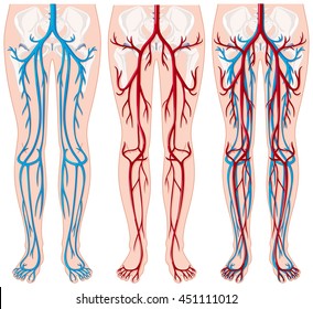
 Blood Vessels Of The Lower Body Stock Vector Illustration
 Figure 4 From The Hemodynamics And Diagnosis Of Venous
 Location Of Venous Reflux In Primary Chronic Venous Disease
 Femoral Vein Wikipedia

 Lower Limb Veins Overview 3d Anatomy Tutorial
 Veins Of Leg Medical Illustration Human Anatomy Drawing
 Finger Foot Human Leg Vein Human Anatomy Png Clipart
 Varicose Veins Maffei Vein Center
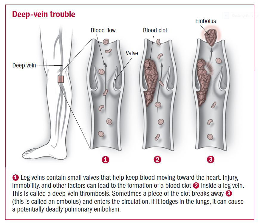 Deep Vein Thrombosis Harvard Health
 Vein Care Specialist Olympia Tacoma Gig Harbor Wa


 History Of Venous Surgery 1 Servier
 How Leg Veins Work Wisconsin Vein Center Varicose Vein
 Varicose Vein Symptoms
 Leg Veins Anatomy Images Stock Photos Vectors Shutterstock
 Blood Vessels Of The Lower Limbs Course Hero

 Venous Drainage Of The Lower Limb Teachmeanatomy
 Glossary Of Terms Medtronic
 Anatomy Springerlink
 Varicose Veins Clinical Gate

 Great Saphenous Vein Anatomy Pictures And Information
 Vein Services Biltmore Cardiology
Belum ada Komentar untuk "Leg Vein Anatomy"
Posting Komentar