Femur Anatomy
The femur or thigh bone is the longest heaviest and strongest bone in the entire human body. Bones of the ankle foot and toes duration.
 Why People Have To Squat Differently The Movement Fix
The femur is found in the thigh.
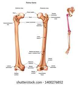
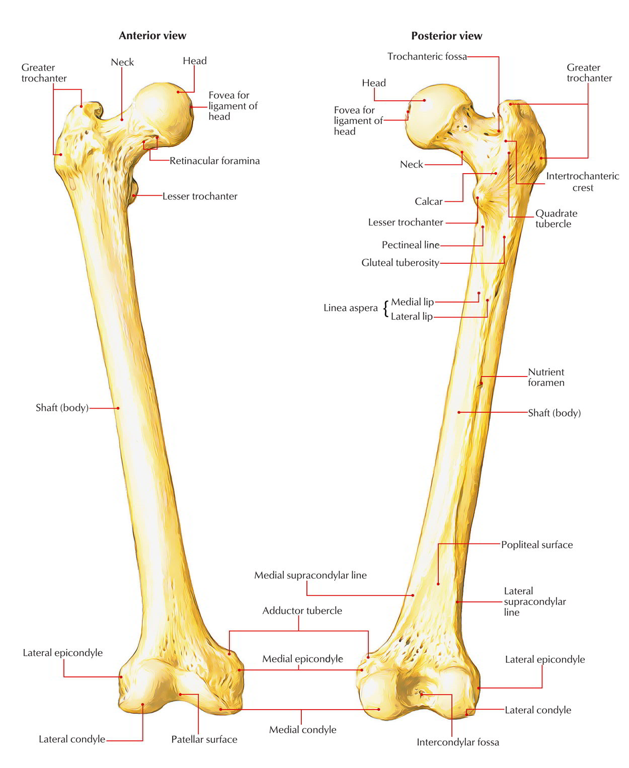
Femur anatomy. Greater trochanter the most lateral palpable projection of bone that originates from. The femur is the primary bone of the leg. The cylindrical shaft is convex forwards.
The femur is the only bone located within the human thigh. It is both the longest and the strongest bone in the human body extending from the hip to read more. All of the bodys weight is supported by the femurs during many activities such as running jumping walking and standing.
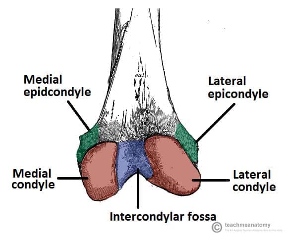
Fractures are the most common condition of the femur. It is both the longest and the strongest bone in the human body extending from the hip to the knee. The upper and bears a rounded head whereas the lower end is widely expanded to from two large condyles.
Femurs or femora ˈ f ɛ m ər ə or thigh bone is the proximal bone of the hindlimb in tetrapod vertebrates and of the human thigh. The femur is the only bone located within the human thigh. The head forms a ball and socket joint with the hip at the acetabulum being held in place by a ligament ligamentum teres femoris within the socket and by strong surrounding ligaments.
The femur ˈ f iː m ər pl. The head is directed medially. The thigh bone is the largest in the body anatomy.
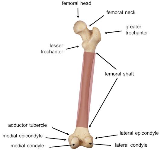
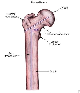
Lesser trochanter smaller than the greater trochanter. By most measures the femur is the strongest bone in the body. Neck connects the head of the femur with the shaft.
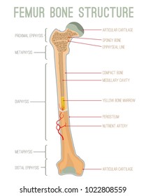
The femur is also called the thigh bone and is the longest and strongest bone of the body. Proximal head articulates with the acetabulum of the pelvis to form the hip joint. It is composed of upper end lower end and a shaft.
Samuel chen 132176 views. The head of the femur articulates with the acetabulum in the pelvic bone forming the hip joint while the distal part of the femur articulates with the tibia and kneecap forming the knee joint. Femur also called thighbone upper bone of the leg or hind leg.
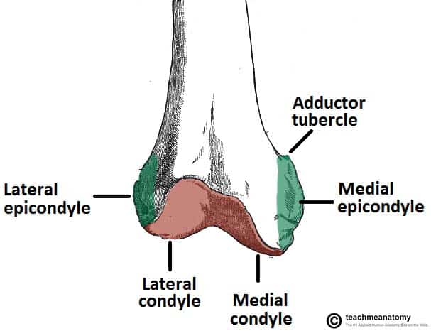
Important features of this bone include the head medial and lateral condyles patellar surface medial and lateral epicondyles and greater and lesser trochanters. It is the largest bone in the body and is. In humans the neck of the femur connects the shaft and head at a 125 angle.
![]() Femur Bone Anatomy Proximal Distal And Shaft Kenhub
Femur Bone Anatomy Proximal Distal And Shaft Kenhub

 The Femur Proximal Distal Shaft Teachmeanatomy
 Femur Anatomy Britannica
 The Femur Human Anatomy
 Easy Notes On Femur Learn In Just 4 Minutes Earth S Lab
 Bones Of The Lower Limb Anatomy And Physiology I
 Femur Radiology Reference Article Radiopaedia Org
![]() Femur Bone Anatomy Proximal Distal And Shaft Kenhub
Femur Bone Anatomy Proximal Distal And Shaft Kenhub
 Hip Fracture Anatomy Causes And Consequences Intechopen
 Royalty Free Femur Stock Images Photos Vectors Shutterstock
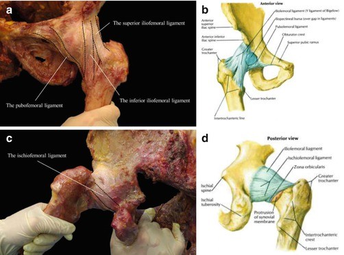 Anatomy Of The Proximal Femur Springerlink
 Femoral Vein Wikipedia
 Proximal Femur Approach Vascular Anatomy Ao Surgery
![]() Femur Bone Anatomy Proximal Distal And Shaft Kenhub
Femur Bone Anatomy Proximal Distal And Shaft Kenhub
 Pediatric Femur Fractures Core Em
Royalty Free Femur Stock Images Photos Vectors Shutterstock
 Osteology Of The Femur

 Femur Anatomy Physiology 251 With Bruder At Chamberlain

 The Femur Proximal Distal Shaft Teachmeanatomy
 Femur Bone Anatomy Landmarks And Muscle Attachments
 Learn Anatomy Online Femur Bone
 The Femur Human Anatomy
 What Is The Anatomy Relative To Intertrochanteric Hip Fractures
 Amazon Com Anatomy Hip Femur Joint Print Sra3 12x18
Belum ada Komentar untuk "Femur Anatomy"
Posting Komentar