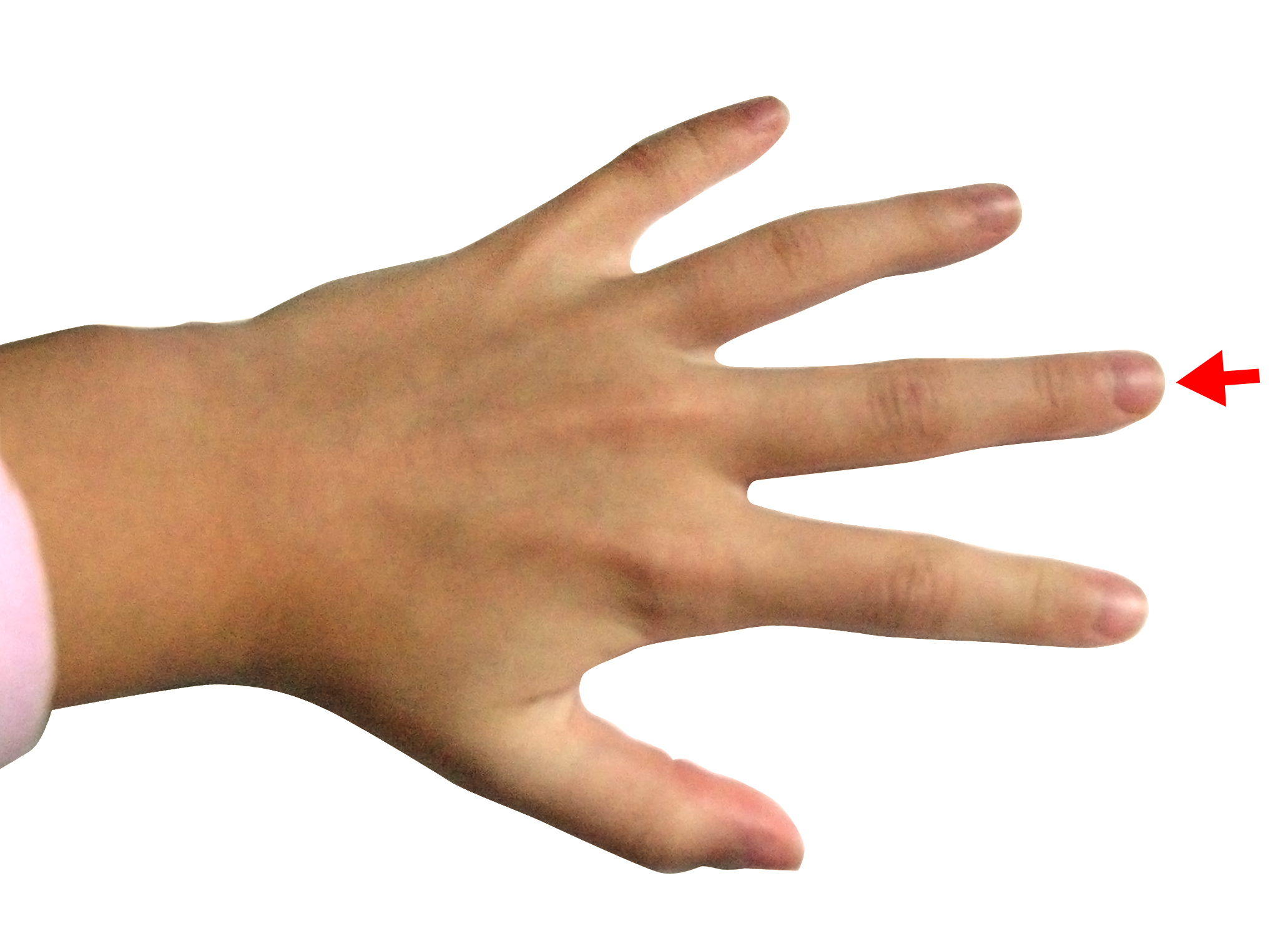
Anatomy Of Index Finger
Finger movement is controlled by muscles in the forearms that pull on finger tendons. This finger often possesses the largest amount of sensitivity and greatest dexterity of any of the fingers.
 Modern Human Thumb And Index Finger Right Hand During Pad
The median nerve innervates the fingers skin.

Anatomy of index finger. There are no muscles in the fingers. The index finger does not contain any muscles but is controlled by muscles in the hand by attachments of tendons to the bones. The extensor digitorum brevis manus is a muscle that originates from the dorsum of the carpus or the distal forearm and inserts into the index finger or less commonly into the long finger.
A digit includes the hand bones but these bones are not separated into individual appendages like a finger. Ligaments connect finger bones and help keep them in place. It works with the extensor digitorum communis to the index finger.
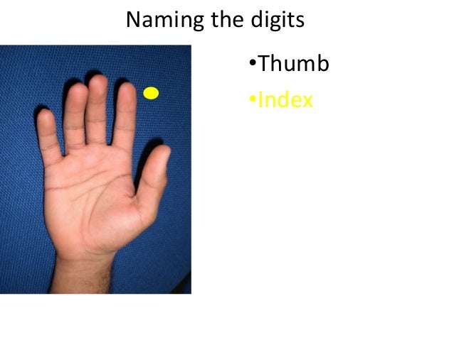
Picture of finger anatomy. The first digit is the thumb followed by index finger middle finger ring finger and little finger or pinkie. Tendons connect muscles to bones.
Each finger has 3 phalanges bones and 3 hinged joints. Fingers have a complex anatomy. Its reported prevalence is from 11 to 47 8 16 17.
Fingers are constructed of ligaments strong supportive tissue connecting bone to bone tendons attachment tissue from muscle to bone and three phalanges bones. A finger is a limb of the human body and a type of digit an organ of manipulation and sensation found in the hands of humans and other primates. Oxygenated blood arrives at the finger through the common palmar artery which extends off of the palmar arch connecting the ulnar and radial arteries.
The extensor indicis extends the index finger while the palmar interosseus adducts it. The thumb has two of each. The pointer finger is the second digit and first finger of the human hand.
The index finger has three phalanges. It has its own muscle belly in the forearm and then as it becomes a tendon it travels through a tough band or retinaculum in the wrist. The index finger is composed of three bones.
Basic anatomy of the finger. It is also called the index finger or the forefinger. The index middle ring and fifth digits have proximal middle and distal phalanges and three hinged joints.
And fingers move by the pull of forearm muscles on the tendons. Anatomy of the fingers the human finger is mainly a bony structure with multiple joints giving it strength and flexibility. Normally humans have five digits the bones of which are termed phalanges on each hand although some people have more or fewer than five due to congenital disorders such as polydactyly or oligodactyly or accidental or medical amputations.
The distal phalanx intermediate phalanx and proximal phalanx. The thumb has a distal and proximal phalanx as well as an interphalangeal and mcp joint. The extensor indicis proprius tendon straightens the index finger.
Distal interphalangeal dip proximal interphalangeal pip and metacarpophalangeal mcp.
Anatomy 101 Finger Joints The Handcare Blog
 Handcare Org Anatomy Photo Gallery
25 Hands Anatomy Mar 30 Flashcards Memorang
 Physical Therapy In Asheville For Trigger Finger And Trigger
 Chronic Hand Pain For 5 Years Talk Tennis
Muscles Of Hands Human Anatomy
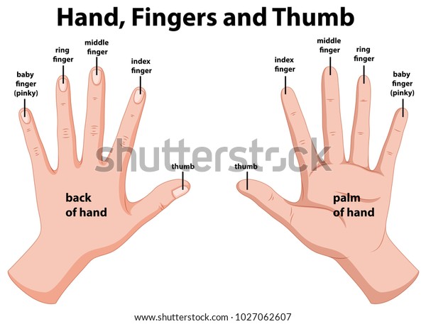 Diagram Of The Fingers Reading Industrial Wiring Diagrams
 Mitten Anatomy The Thumb Gusset Interweave

Degloving Injury To The Left Index Finger With Surgical Flap
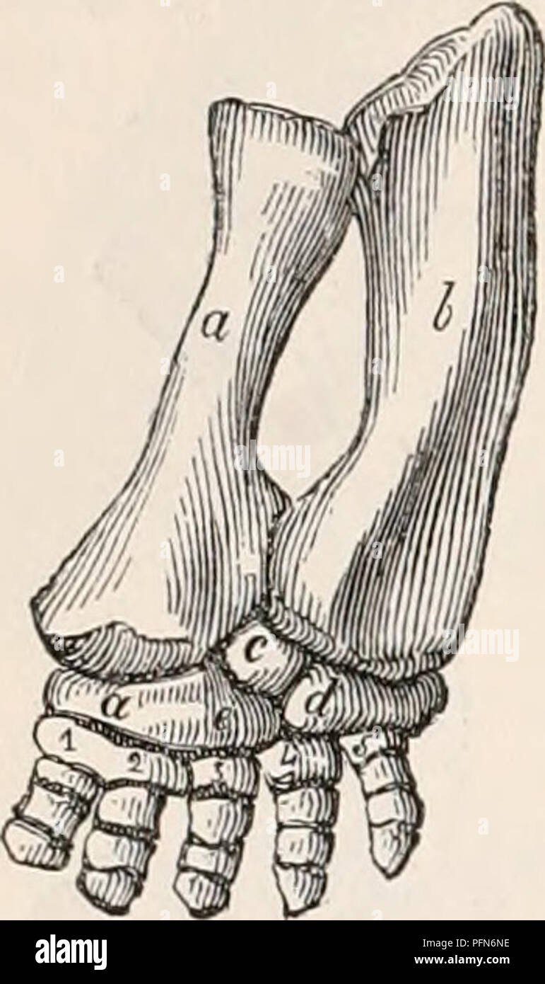 The Cyclopaedia Of Anatomy And Physiology Anatomy
 Injuries Of The Left Index Finger With Surgical Repairs
Pollicization Boston Children S Hospital
Patient Education Concord Orthopaedics
 Left Index Finger A Crucial Anatomy For Ge14 New Straits
 The Muscles And Fasciae Of The Forearm Human Anatomy
 Hand Finger Anatomy Hand Anatomy Hand Surgery Human Anatomy
 Anatomy Bones Of The Index Finger
 Our Index Finger Pointing To The Creator Answers In Genesis
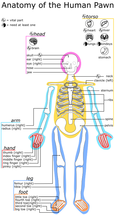



Belum ada Komentar untuk "Anatomy Of Index Finger"
Posting Komentar