Female Anatomy Pregnancy
Your period has stopped and you may have nausea breast tenderness and swelling frequent urination and fatigue. During these final weeks your baby continues to grow and develop.
 Female Reproductive Anatomy Pregnancy Birth Poster 12x17
At this point your uterus has begun to grow and become more egg shaped.

Female anatomy pregnancy. If there is no pregnancy then the uterus sheds this lining menstruation. A woman is born with 200000 eggs but by the time she reaches puberty that number has dwindled to 400 or so. The male and female reproductive systems provide essential components for achieving pregnancy.
It starts at week 28 of your pregnancy and ends with the birth of your baby. The system is designed to. Everything from belly size to heartbeat speed will change over the 9 months leading up to childbirth.
The male reproductive system produces and deposits sperm. The portion of the uterus superior to the opening of the uterine tubes is called the fundus. When you are between 39 and 41 weeks your pregnancy is considered full term and your baby is ready to be born.
The 3d pregnant female morph model is a derivative of the 3d female integumentary system with geometry carefully crafted to provide resolution in key areas for high quality rendering with excessive and superfluous geometry removed to increase functionality of the product. Its average size is approximately 5 cm wide by 7 cm long approximately 2 in by 3 in when a female is not pregnant. The uterus grows a lining each month in preparation for pregnancy.
The ovaries where eggs are stored and released usually one each month connect to the uterus via the fallopian tubes. Your body at 6 7 weeks of pregnancy when you are between 6 and 7 weeks pregnant you may be experiencing the early signs of pregnancy. The heart begins to beat in the developing offspring.
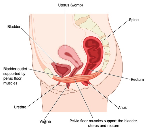
The uterus or womb is a hollow pear shaped organ. Pregnancy is a time of great physical and emotional change for women. It has three sections.
The ovaries also release the female sex hormones estrogen and progesterone. Two fallopian tubes one on each side stretch from the ovaries to the uterus. It can expand up to 50 cm in length during pregnancy.
It produces the female egg cells necessary for reproduction called the ova or oocytes. The female reproductive system is designed to carry out several functions. It signals the corpus luteum to continue producing estrogen and progesterone to maintain the pregnancy.
Lets focus on the third trimester. The female reproductive system produces eggs and protects and nourishes a growing baby until birth.
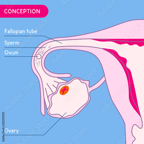 Fecundation Female Reproductive System Human Anatomy
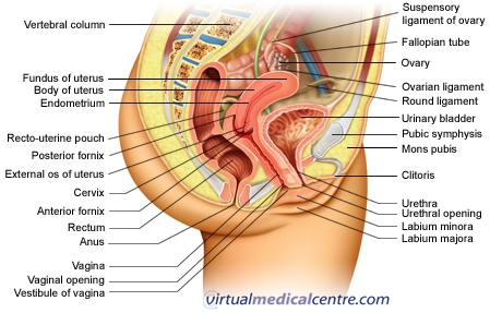 Ectopic Pregnancy Healthengine Blog
![]() Reproductive System Female Anatomy Uterus Cervix Fallopian
Reproductive System Female Anatomy Uterus Cervix Fallopian
 Clip Art Vector Normal Pregnant Female Anatomy Stock Eps
 36 Part Multi Torso Anatomy Model Male Female Sexless And Pregnancy Configuration
 Supreme 4d Master Human Anatomy Model Female Transparent
 Details About Human Female Pelvic Section Pregnancy Anatomical Model Medical Pelvis Anatomy
 Female Pelvis With Uterus In The Ninth Month Pregnancy Model Female Anatomy Pelvis With Baby Model Buy Female Pelvis With Uterus Female Anatomy

 Female Pelvic Anatomy Early In First Pregnancy Medical Exhibit
 Anatomy Of A Pregnant Woman Vector Image 1863663

 Female Reproductive System Isolated Human Anatomy
 Reproductive System Female Anatomy Uterus Cervix Fallopian Tube Pregnancy Test Icon Healthcare Medical Service Logo Medicine Symbol Concept Seamless
 How Your Body Changes In Pregnancy Video Babycentre Uk
 Watercolor Painted Silhouette Of Side View Pregnancy Process
 Pregnancy Birth Chart 22x28 Clinicalposters
 Details About Classic Pregnancy 8 Model Series Set Anatomy Female Pregnancy Models School New
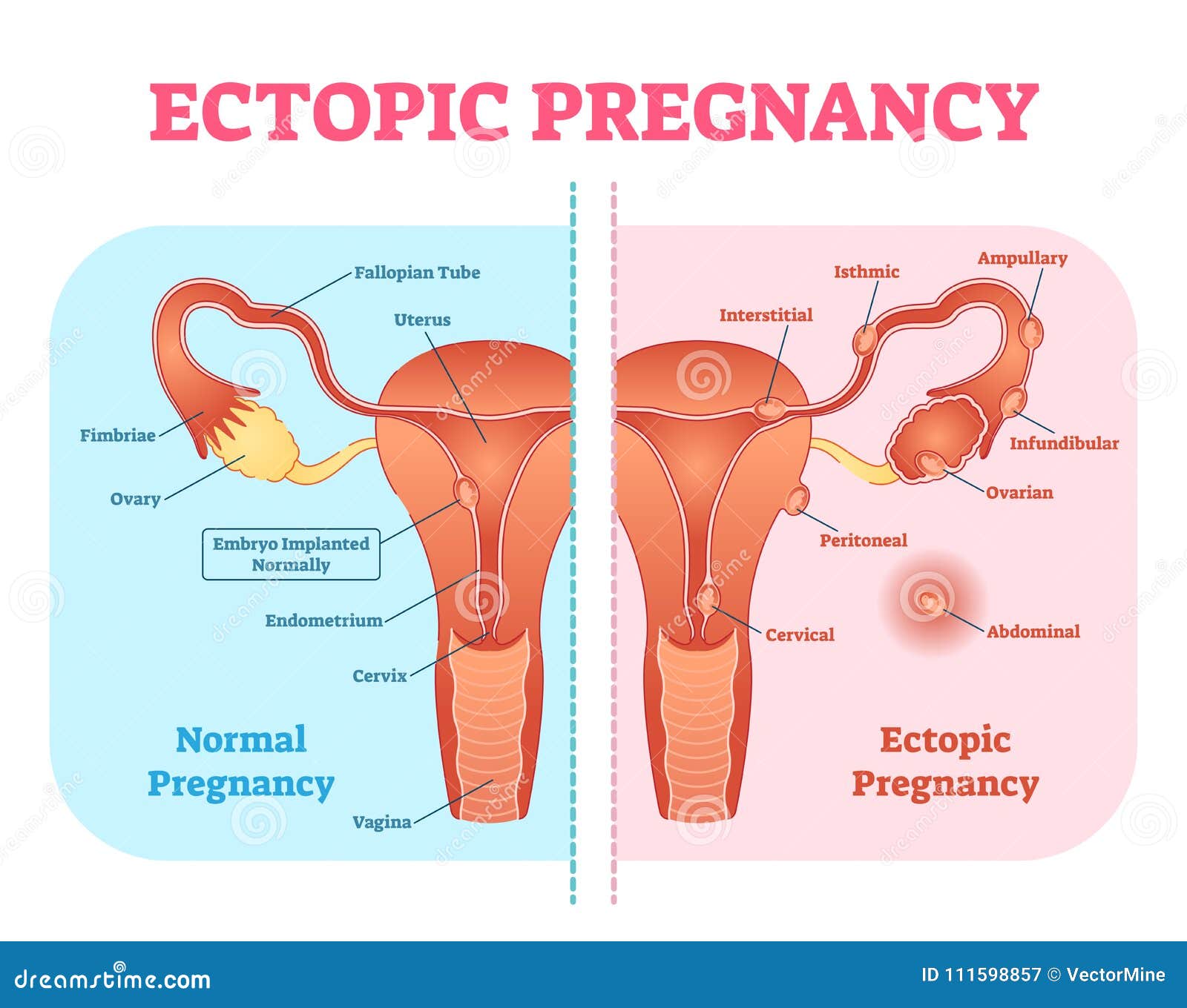 Ectopic Pregnancy Or Tubal Pregnancy Medical Diagram With
 Supreme Female Anatomy Model Clear
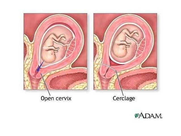 Anatomical Changes In Pregnancy

Belum ada Komentar untuk "Female Anatomy Pregnancy"
Posting Komentar