Phrenic Nerve Anatomy
The phrenic nerve can be marked by a line connecting these two points. Understanding the anatomy of the phrenic nerves is essential for surgeons.
 Instant Anatomy Diagram
Descends anteriorly along the right lung root.
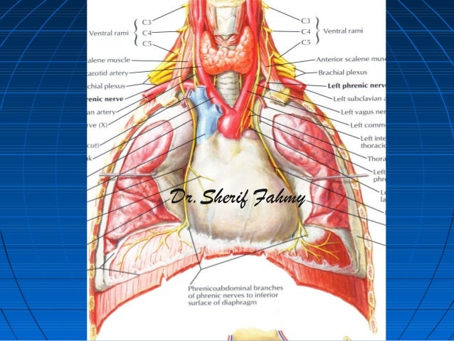
Phrenic nerve anatomy. 1st point can be labelled 35 cm at the level of the thyroid cartilage from the midsagittal plane. It crosses the arch lateral to the superior intercostal vein and in front of the vagus nerve and then runs laterally down the pericardium over the left ventricle towards the apex of the heart. The left phrenic nerve runs laterally to the aortic arch and initial parts of the left subclavian artery and the left common carotid artery.
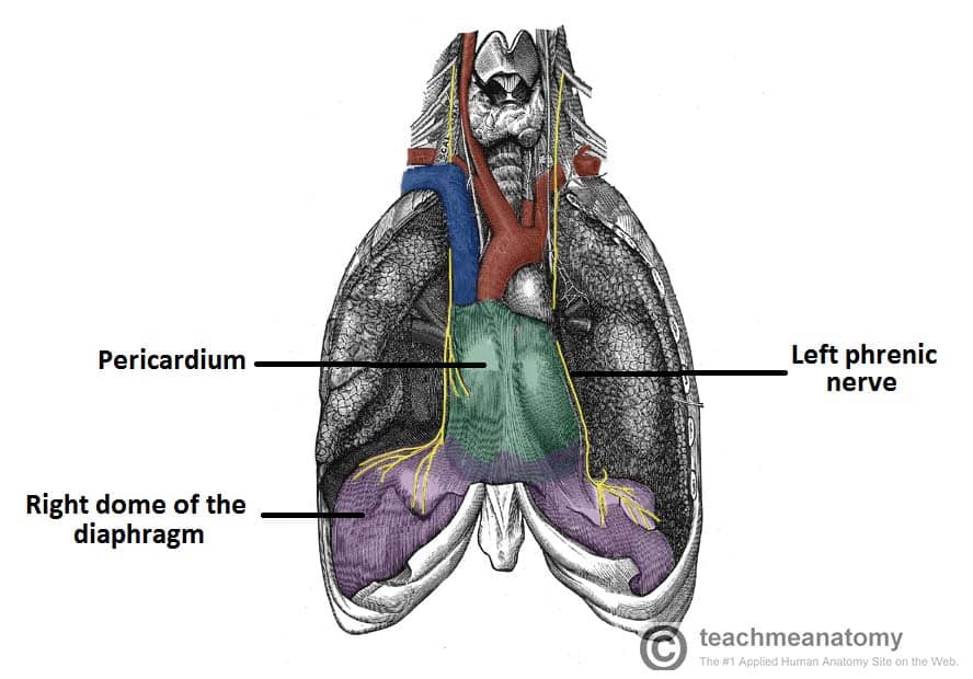
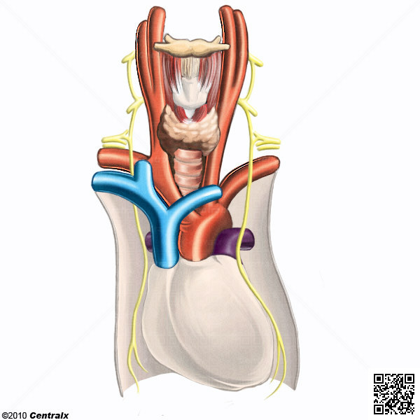
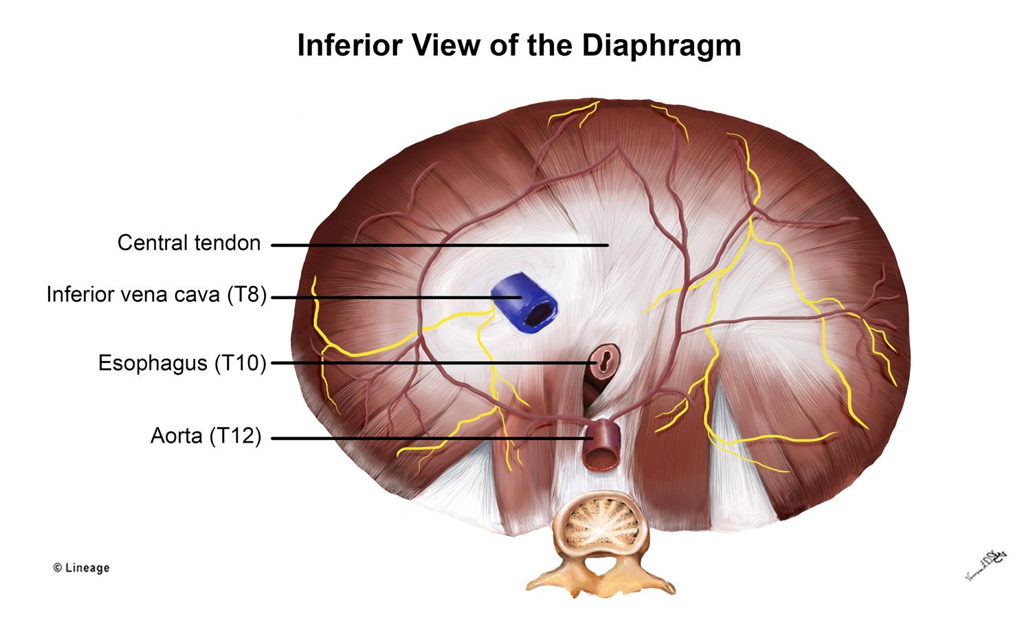
Courses along the pericardium of the right atrium of the heart. Then the phrenic nerves go down. It provides complete motor innervation to the diaphragm and sensation to the central tendon aspect of the diaphragm.
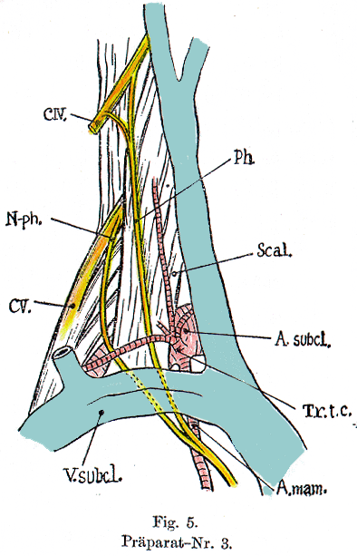
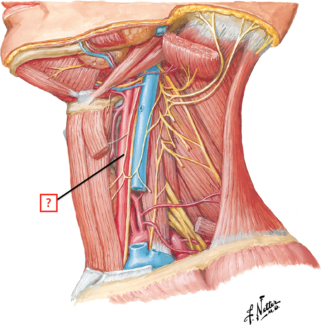
At the level of the mediastinum the phrenic nerve is found alongside the anterior scalene muscle shielded in the fibrous connective tissue that surrounds the vascular structures of the neck more commonly known as the carotid sheath. The phrenic nerve is a mixed motorsensory nerve that courses through the neck and thorax to innervate the diaphragm. Conditions associated with phrenic nerve function or dysfunction can range.
Phrenic nerve anatomy definition phrenic nerve arises in neck region from the anterior rami of the 3rd 4th and 5th cervical spinal nerves. The phrenic nerve has sensory motor and sympathetic functions. From there the two phrenic nerves follow different directions one more.
The left phrenic nerve has the left common carotid and left subclavian arteries that arise from the arch of the aorta to its medial side. 2nd point is at the medial end of the clavicle. In the upper chest the right phrenic nerve lies lateral to the right brachiocephalic vein and the superior vena cava.
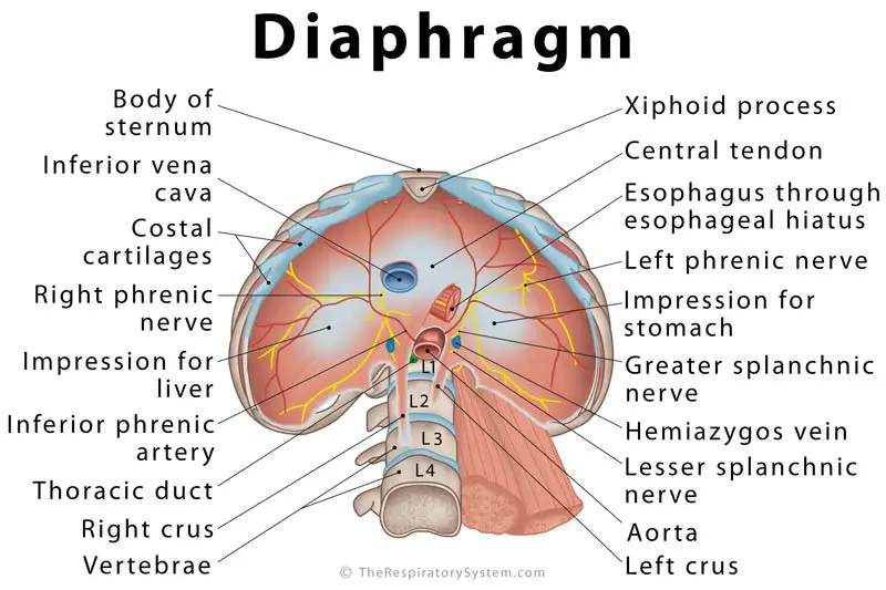
Pierces the diaphragm at the inferior. The anatomy of the phrenic nerve anatomy. Right phrenic nerve passes anteriorly over the lateral part of the right subclavian artery.
Dr yuranga weerakkody and dr henry knipe et al. Enters the thorax via the superior thoracic aperture. The phrenic nerve originates from the anterior rami of the c3 through c5 nerve roots and consists of motor sensory and sympathetic nerve fibers.
Phrenic nerve roots are c3c4c5 but mainly from the fourth cervical spinal segment root and recive contribution from third and forth cervical nerves.

 The Phrenic Nerve Anatomical Course Functions
 Which Artery Accompanies The Phrenic Nerve In The Mediastinum
 Left Atrial Anatomy Relevant To Catheter Ablation Figure 12
 Diaphragm Definition Location Anatomy Function Diagram
:max_bytes(150000):strip_icc()/iStock-464631030-098208378b8340c1b2cf7eb6290a80a1.jpg) Phrenic Nerve Anatomy Function And Treatment
Phrenic Nerve Anatomy Function And Treatment
Phrenic Nerve Vagus Nerve Anatomy Of He Thorax
Right Phrenic Nerve
 Phrenic Nerve Diaphragm And Pericardium Innervation
 Human Anatomy Lab 21 Step 7
 Anatomy Atlases Illustrated Encyclopedia Of Human Anatomic
 Supplement To Motor Axon Guidance Of The Mammalian Trochlear
 Phrenic Nerve
 Phrenic Nerve Damage Causes And Treatments Avery
 Phrenic Nerve An Overview Sciencedirect Topics
 Lung And Posterior Mediastinum Anatomy Test 2 Flashcards
 Phrenic Nerve Block Springerlink
 Photograph Shows The Variations Of The Phrenic Nerve Ac
 Anatomy Atlases Illustrated Encyclopedia Of Human Anatomic
 Diaphragm Respiratory Medbullets Step 1
 Phrenic Nerve An Overview Sciencedirect Topics
 The Phrenic Nerve
 Left Phrenic Nerve Anatomy Relative To The Coronary Venous System
 Anatomy Pericardium External Heart And Phrenic Nerves Week
Belum ada Komentar untuk "Phrenic Nerve Anatomy"
Posting Komentar