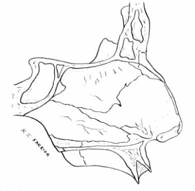
Vomer Anatomy
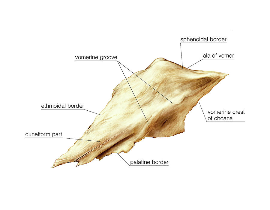
The anterior border is the longest and slopes downward and forward. 174 are marked by small furrows for blood vessels.
 Nasal Anatomy Embryology Skin And Soft Tissues Blood
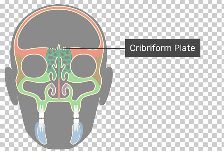
The septum separates the nasal cavity into two halves called nasal fossae.
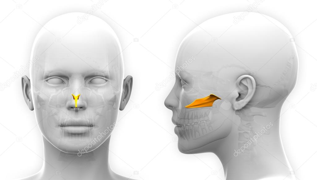
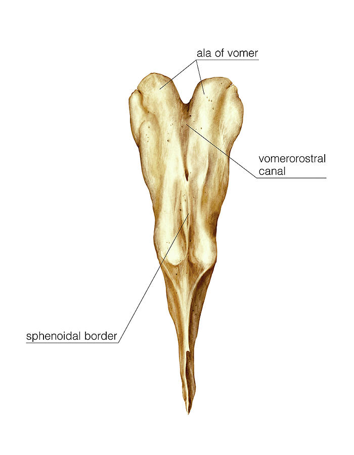
Vomer anatomy. With the help of these wings the vomer attaches to the inferior surface of the body of the sphenoid bone. The vomer is one of the facial bones and forms the postero inferior part of the bony nasal septum. The vomer ˈ v oʊ m ər is one of the unpaired facial bones of the skull.
The vomer is situated in the median plane but its anterior portion is frequently bent to one or other side. Vomer bone l plowshare a thin plow shaped bone. It has two surfaces and four borders.
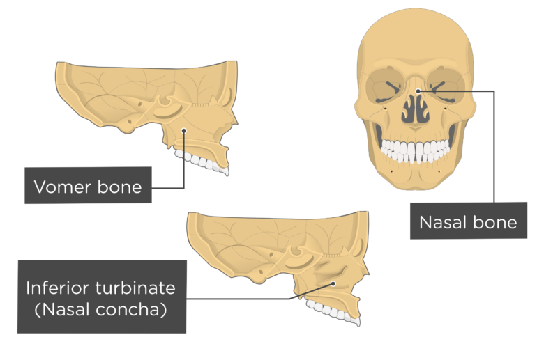
The vomer articulates with the following bones. In skull cavity is formed by the vomer and the nasal lachrymal and turbinate bones. The vomer has 4 borders.
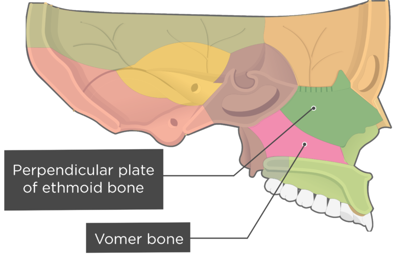
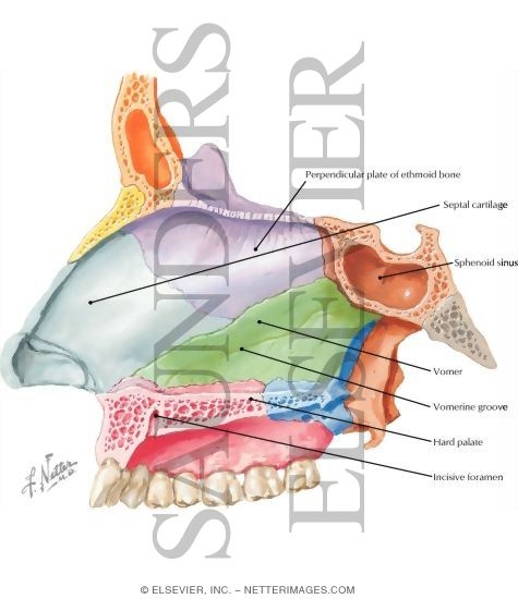
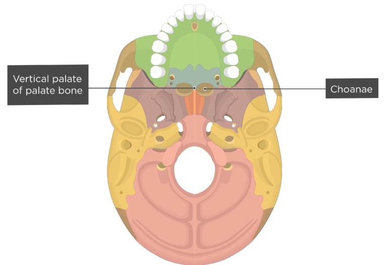
The posterior border is free concave and separates the choanæ. The vertical plate of the vomer bone articulates with the perpendicular plate of the ethmoid bone to form the bony nasal septum. Articulates with the median nasal crests of the maxilla and palatine bones.
It is thick and bifid above thin below. Many mammals such as the dog have a sagittal crest down the centre of the skull. It is a thin flat bone that is trapezoidal in shape with two surfaces that are obliquely grooved by the sphenopalatine nasopalatine vessels and nerves.
Its lower half is grooved for the inferior margin of the septal cartilage of the nose. In infants the sutures joints between the various skull elements are loose but with age they fuse together. It is unpaired and lies in the midline between the two nasal cavities.
Short concave border the. The vomer is a single facial bone that takes a form of a plate and its superior border gives two extensions called wings. Thickest border with laterally projecting alae which articulate with the rostrum.
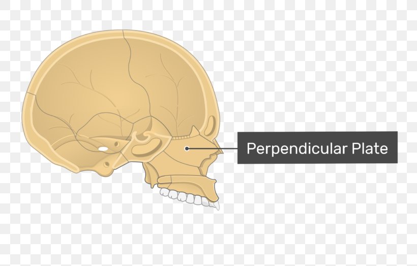
Its upper half is fused with the perpendicular plate of the ethmoid. It is thin somewhat quadrilateral in shape and forms the hinder and lower part of the nasal septum fig. It is located in the midsagittal line and articulates with the sphenoid the ethmoid the left and right palatine bones and the left and right maxillary bones.
Longest border which articulates with.
 Anatomy 2 Year Midterm Test Flashcards Quizlet
 Nasal Vomer And Inferior Turbinate Concha Bones Anatomy
 Vomer
Vomer
 Perpendicular Plate Of Ethmoid Bone A D A M Interactive
Ala Of Vomer
 Nasal Vomer And Inferior Turbinate Concha Bones Anatomy
 Bones Of The Cranium Course Hero

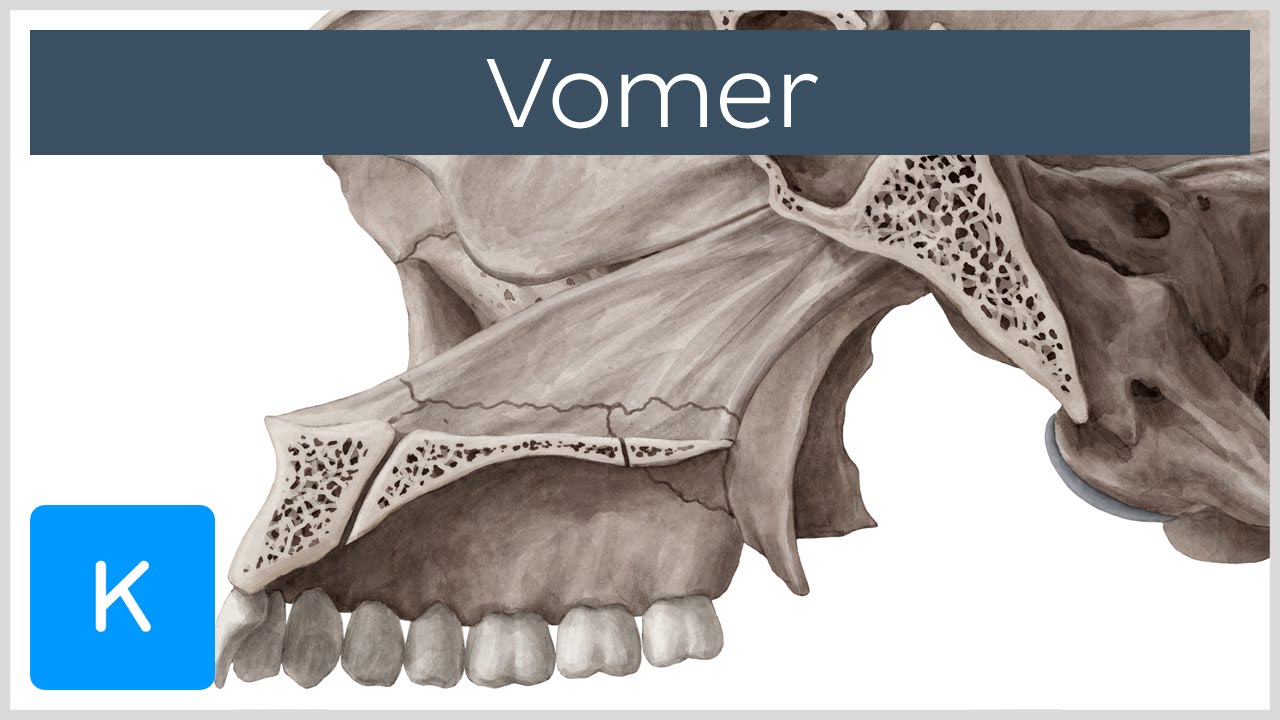
![]() Vomer Anatomy Structure And Articulations Kenhub
Vomer Anatomy Structure And Articulations Kenhub
 Ethmoid Bone Ethmoid Sinus Vomer Anatomy Png Clipart
 Image Result For Vomer Bone Bones Anatomy Veterinary
 Vomer Wikipedia
 Anatomy Of Articulation And Resonation Csdi 4037 Ppt Video
 The Skull Anatomy And Physiology I
 Vomer
Vomer
 Brain Anatomy Goodman Campbell Brain And Spine

 Vomer
Vomer
 7 3 The Skull Anatomy Physiology

 Nasal Vomer And Inferior Turbinate Concha Bones Anatomy
 Nasal Vomer And Inferior Turbinate Concha Bones Anatomy
Female Vomer Skull Anatomy Isolated On White Stock Photo
 The Palatine Bone Human Anatomy
 Seer Training Axial Skeleton 80 Bones
 Vomer Bone Definition Location Human Anatomy Kenhub
![]() Vomer Anatomy Structure And Articulations Kenhub
Vomer Anatomy Structure And Articulations Kenhub
Belum ada Komentar untuk "Vomer Anatomy"
Posting Komentar