Anatomy Of The Foot And Ankle
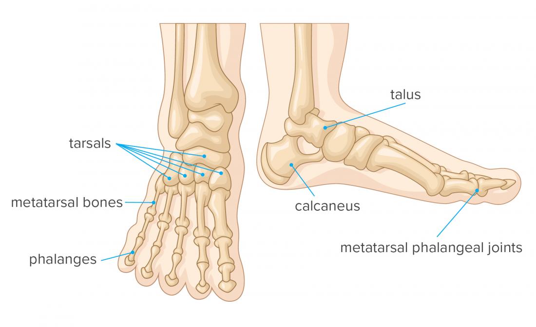
The midfoot is a pyramid like collection of bones that form the arches of the feet. The last two together are called the lower ankle joint.
The talus bone supports the leg bones tibia and fibula forming the ankle.

Anatomy of the foot and ankle. Hardcover 9590 95. The ankle joint talocrural joint is formed where the distal end of the leg meets the foot. By kelikian md armen s and sarrafian md facs shahan k.
Use our anatomy tools to learn about bones joints ligaments and muscles of the foot and ankle. 90 to rent 13970 to buy. The ankle joint allows up and down movement of the foot.
These muscles allow the ankle to bend downward and outward. These include the three cuneiform bones the cuboid bone and the navicular bone. Ebraheims educational animated video describes anatomical structures of the foot and ankle the bony anatomy the joints ligaments and the compartments in a simple and easy way.
Footeducation is committed to helping educate patients about foot and ankle conditions by providing high quality accurate and easy to understand information. The peroneal muscles peroneus longus and peroneus brevis on the outside edge of the ankle and foot. Upper ankle joint tibiotarsal talocalcaneonavicular and subtalar joints.
The bones of the foot and ankle begin with the ankle joint itself. The ankle joint is formed where the talus the uppermost bone in the foot and the tibia shin meet. The hindfoot forms the heel and ankle.
The largest and strongest tendon of the foot is the achilles tendon which extends from the calf muscle to the heel. Sarrafians anatomy of the foot and ankle. Foot ankle anatomy muscles tendons and ligaments.
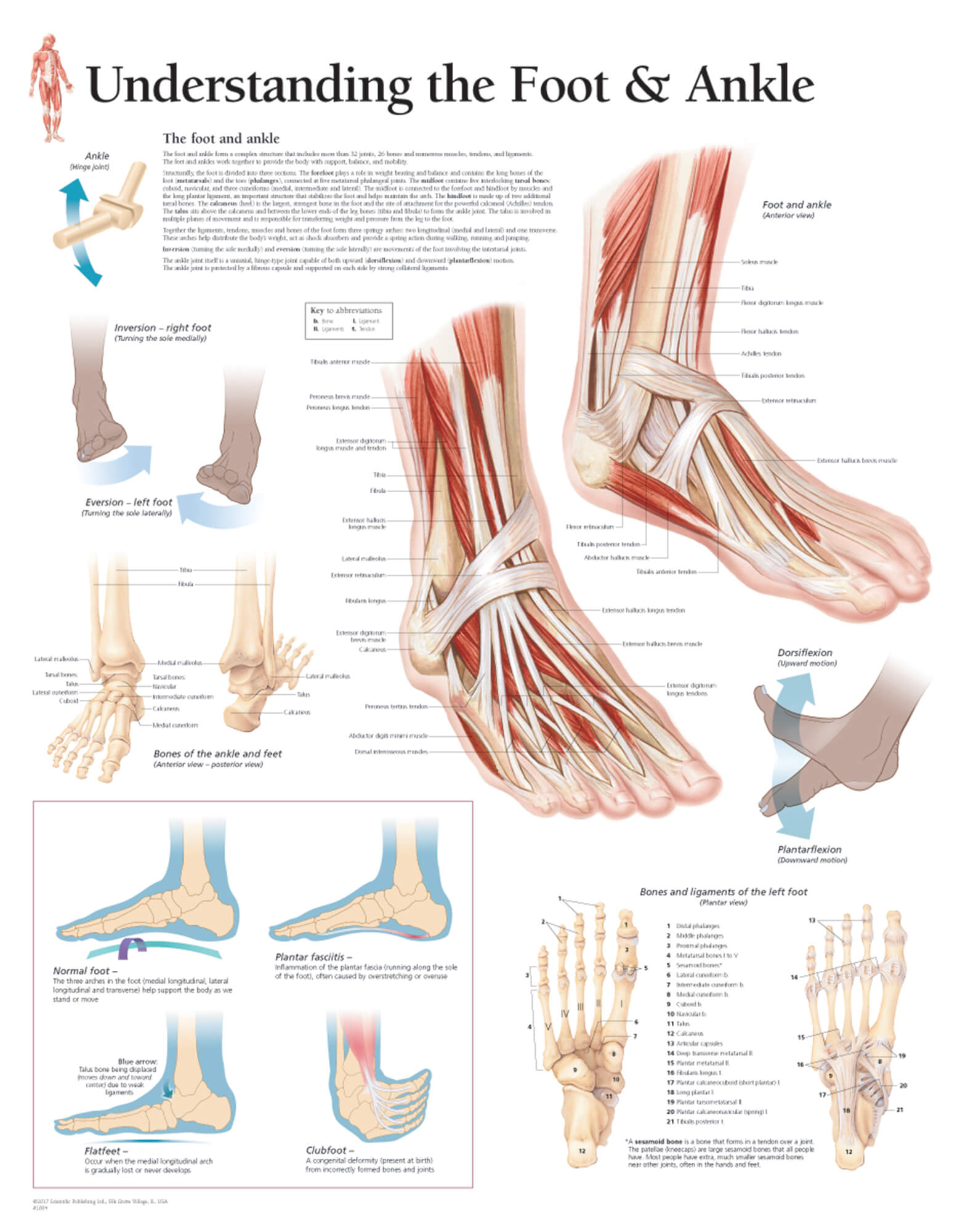
Foot and ankle anatomy is quite complex. These all work together to bear weight allow movement and provide a stable base for us to stand and move on. There are also multiple muscles in the ankle that can be strained as follows.
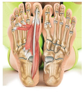
There are elastic tissues tendons in the foot that connect the muscles to the bones and joints. Free shipping by amazon. It is made up of three joints.
The subtalar joint sits below the ankle joint and allows side to side motion of the foot. Get it as soon as mon jul 29. The ankle joint also known as the talocrural joint allows dorsiflexion and plantar flexion of the foot.
50 out of 5 stars 6. Major muscles of the ankle. The foot consists of thirty three bones twenty six joints and over a hundred muscles ligaments and tendons.
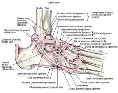
Lateral side of the ankle joint capsule. Its strength and joint function facilitate running jumping walking up stairs and raising the body onto the toes.
 Foot Anatomy Spokane Valley Wa Foot Doctor
 Foot And Ankle Chart 20x26 Clinicalposters
Anatomy And Injuries Of The Foot And Ankle Chart 20x26
 Foot Ankle Anatomy Pictures Function Treatment Sprain Pain
.jpg) Foot Anatomy Spokane Valley Wa Foot Doctor
Foot Anatomy Spokane Valley Wa Foot Doctor
 Muscles Of The Foot Laminated Anatomy Chart
 1 Bony Anatomy Of The Foot And Ankle Download Scientific
 Test Anatomy Of Foot Ankle And Lower Leg Quizlet
:background_color(FFFFFF):format(jpeg)/images/library/11041/anatomy-ankle-joint_english.jpg) Ankle And Foot Anatomy Bones Joints Muscles Kenhub
Ankle And Foot Anatomy Bones Joints Muscles Kenhub
 Foot And Ankle Anatomy Allen Tx Foot Doctor
 Foot And Ankle Anatomy Bones Muscles Ligaments Tendons
Foot And Ankle Anatomy San Diego Coronado La Jolla Del
 Illustrated Anatomy Of The Foot A The Cuneiforms Cuboid
 Anatomy Of The Foot North Arkansas Podiatry
 Mr Miles Callahan Anatomy Of The Foot And Ankle
 Ankle Joint Anatomy Overview Lateral Ligament Anatomy And
Rheumatoid Arthritis Of The Foot And Ankle Orthoinfo Aaos
 Foot Bones Anatomy Conditions And More
 Topographic Anatomy Of The Foot And Ankle
 Axis Scientific Foot And Ankle Joint Section Anatomy Model


Belum ada Komentar untuk "Anatomy Of The Foot And Ankle"
Posting Komentar