Horse Anatomy Stifle
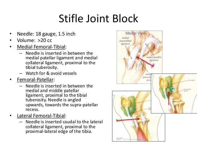
Inside the equine stifle. The femoropatellar joint medial femorotibial joint and lateral femorotibial joint.
The mother of all joints.
Horse anatomy stifle. Wrapping your mind around the anatomy of the stifle joint can take a few minutes but its important for understanding the diagnostic and treatment challenges it presents. Stifle anatomy radiograph patella femur the equine stifle corresponds to the human knee. The equine stifle consists of three compartments.
Although horsemen refer to the stifle as if it were a single joint its actually a three for one deal with lots of extras thrown in. The horses stifle is akin to a human knee and it usually bends forward. It is the equivalent of the human knee and is often the largest synovial joint in the animals body.
Anatomy the stifle is the equivalent of the human knee and it is the largest most complex joint in the horse. Femoropatellar medial femorotibial and lateral femorotibial. A horse with a locked stifle will likely hold its hind leg stiff and straight unable to unlock the joint.
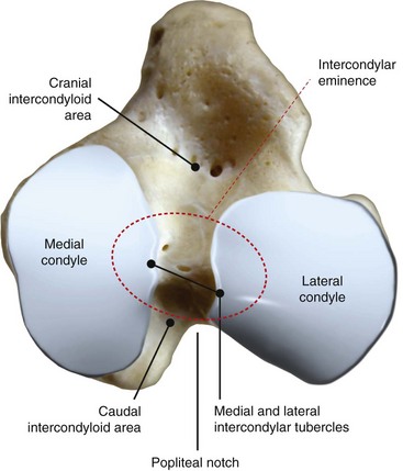
The stifle joint joins three bones. 13 percent had osteochondrosis dissecans ocd lesions while a small percentage of horses showed either osteoarthritis 3 percent fractures 4 percent epiphysitis 1 percent or unknown causes 13 percent. Instead there is an intertubercular bursa.
Communication of the femoropatellar and medial femorotibial joints has been found 60 to 70 of the time although inflammation anatomic variation and unidirectional flow affect this communication. This study showed 15 percent had upward fixation of the patella. The bones that make up the stifle are the femur thigh tibia shin and patella kneecap.
The bones that make up the stifle are the femur thigh tibia shin and patella kneecap. The radiograph at left credit vetwerx is a lateral view of the stifle showing the knee cap or patella and the femur. Stifle joint the stifle is the equivalent of the human knee and it is the largest most complex joint in the horse.
The femur patella and tibia. The joint consists of three smaller ones. The stifle joint often simply stifle is a complex joint in the hind limbs of quadruped mammals such as the sheep horse or dog.
This bursa lies between the humeral tubercles cushioning the bicipital tendon but does not communicate with the cavity of the shoulder joint. Observe your horse to see if it holds its leg taut and if it drags the toes of its hoof on the ground behind it. In the horse there is no sheath surrounding the bicipital tendon.
The stifle lifts the leg upward and forward making it critical to moving and athletic pursuits. As the leg moves the patella rides up and down the trochlear ridges of the femur in the femoropatellar joint. The stifle lifts the leg upward and forward so its pretty critical to moving.
 138 Best Equine System Skeletal Joint Images Horses
Aec Client Education Upward Patellar Fixation
 Horse Leg Bone Anatomy If You Look At The Horse From
 The Equine Tarsus Hock Vet Physio Phyle
 Horse Anatomy I Mikki Senkarik
 Regional Anesthesia In Equine Lameness Musculoskeletal
Traumatic Stifle Injuries In The Horse Equitrader
 The Stifle Joint Of The Horse Mackinnon
 The Anatomy Of The Horse A Dissection Guide Horses
 Equine Stifle An Overview Sciencedirect Topics
 Locking Stifles Henderson Equine Clinic
Common Joint Diseases Of Horses
 Equine Stay Apparatus Hindlimb 3d Veterinary Anatomy Learning Ivala
 The Anatomy Of Dressage Horse Hindquarters Expert Advice
 Horse Selection Animal Food Sciences
 Anatomy Of The Horse Osteology
 Parts Of A Horse Teaching Aid Osu Extension Catalog
 Equine Stifle In Parker Berthoud Boulder Co Vetwerx Equine
Horse Equine Wound Injury Bandage Step Ahead Structure
How To Perform Arthrocentesis Of The Compartments Of The
 The Anatomy Of The Horse A Dissection Guide Horses Viii
How To Perform Arthrocentesis Of The Compartments Of The
 Equine Stifle An Overview Sciencedirect Topics


Belum ada Komentar untuk "Horse Anatomy Stifle"
Posting Komentar