Anatomy Of Bones
Red bone marrow is where all new red blood cells white blood cells and platelets are made. Types long bones are characterized by a shaft the diaphysis that is much longer than its width.
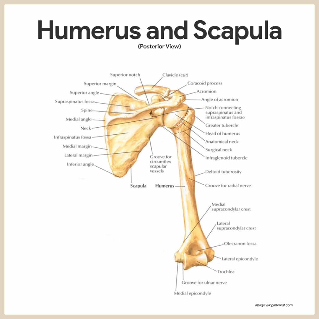 Skeletal System Anatomy And Physiology Nurseslabs
The four general categories of bones are long bones short bones flat bones and irregular bones.
Anatomy of bones. He talks about the anatomy of the skeletal system including the flat short and irregular bones and their individual arrangements of compact and spongy bone. Gross anatomy of bone. Short bones include the carpal and tarsal bones patellae and sesamoid bones.
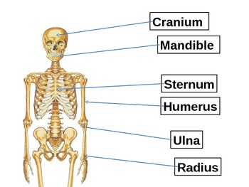
Long bones include the clavicles humeri radii ulnae metacarpals femurs tibiae fibulae metatarsals and phalanges. There are two types of bone marrow. Intertubercle sulcus or bicipital groo parasagittal vs midsagittal para is parallel mid is a plane that cuts them into equal par.
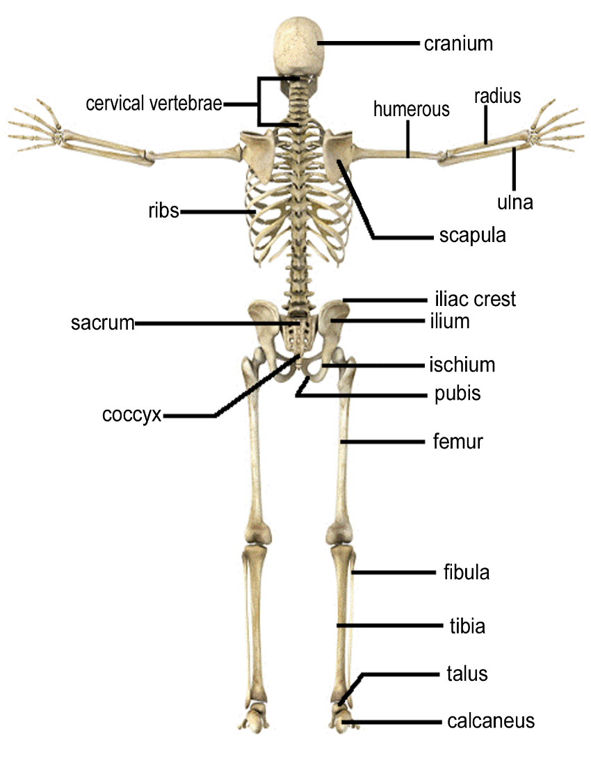
Anatomy of the typical long bone as well as microscopic anatomy of bone tissue. The periosteum contains blood vessels nerves and lymphatic vessels that nourish compact bone. The axial skeleton runs along the bodys midline axis and is made up of 80 bones in the following regions.
Flat bones are thin and generally curved with two parallel layers of compact bones sandwiching. These are long bones short bones flat bones irregular. Gross anatomy of bone.
Short bones are roughly cube shaped and have only a thin layer of compact bone surrounding. Platelets are small pieces of cells that help you stop bleeding when you get a cut. The diaphysis and the epiphysis.
Bones can be described in many ways and one of the easiest ways to categorize them is by their shape. There are five main shapes of bones. The outer surface of the bone is covered with a fibrous membrane called the periosteum peri around or surrounding.
The diaphysis is the tubular shaft that runs between the proximal and distal ends of the bone. A long bone has two parts. The inside of your bones are filled with a soft tissue called marrow.
Creates foramen supras greaterlesser tubercle. The structure of a long bone allows for the best visualization of all of the parts of a bone figure 1. Para is parallel mid is a plane that cuts them into equal par first bone in ossification 5 weeks in fetus.
Tendons and ligaments also attach to bones at the periosteum.
 Anatomy Gross Anatomy Physiology Cells Cytology Cell
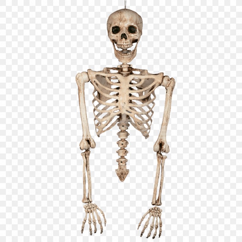 Human Skeleton Bone Torso Human Body Png 908x908px

 Bones Of The Lower Limb Anatomy And Physiology
 Why Crossfit Coaches Need Anatomy Bones Muscles And
 Vector Illustration Female Pelvis Bone Anatomy Eps
 Infographic Diagram Of Human Skeleton Lower Limb Anatomy Bone
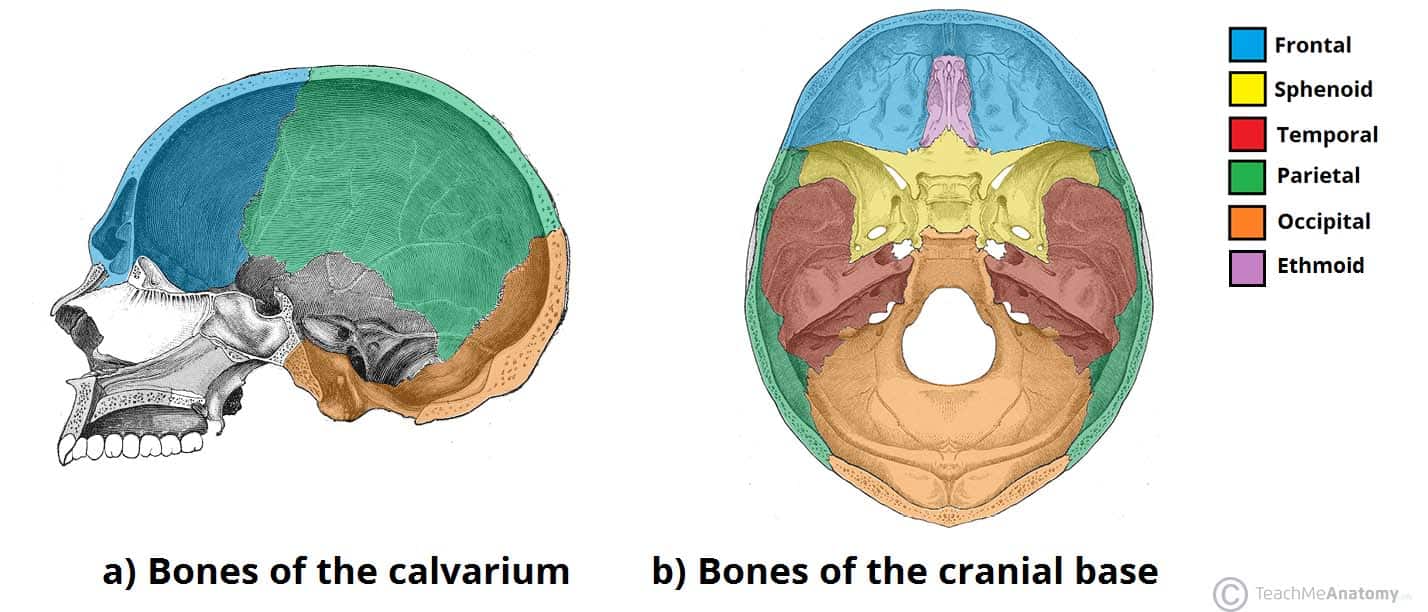 Bones Of The Skull Structure Fractures Teachmeanatomy
 Skeleton Bones Rear Medical Anatomy Bones
 The Human Skeleton Bones Structure Function Teachpe Com
 Us 7 32 41 Off Xr607 Canvas Painting Wall Art Picture Human Skeletal Anatomy Bones Of The Body Poster Print Body Map Pictures For Medical Educa In
 Vintage Anatomy Print Bones Of Foot
 Monmed Medical Skeleton Model Small Skeleton Model Human Skeleton Model For Anatomy Art Halloween Decor 17 Inch
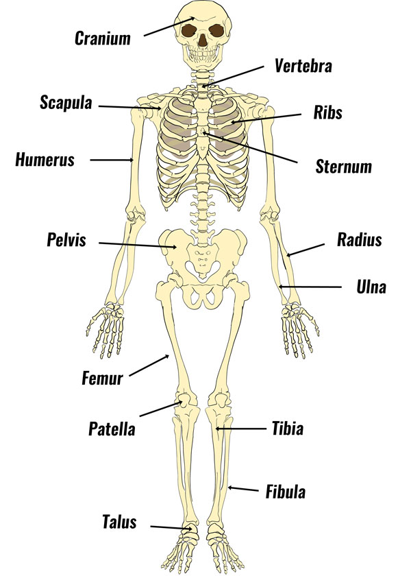 The Human Skeleton Bones Structure Function Teachpe Com
 Skeletal System Anatomy And Physiology Nurseslabs
 Anatomy Of Hand Bones Stock Vector Illustration Of Health
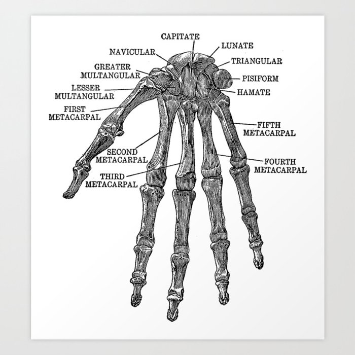 Bones Of The Human Hand Anatomy Skeleton Art Print By Wandastar
 Human Skeleton Parts Functions Diagram Facts Britannica
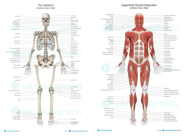 Welcome To Ms Stephens Anatomy And Physiology And
Anatomy Gross Anatomy Physiology Cells Cytology Cell
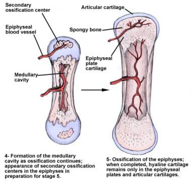 Osteology Bone Anatomy Overview Gross Anatomy Overview

Belum ada Komentar untuk "Anatomy Of Bones"
Posting Komentar