Female Hip Anatomy
The femur has a ball shaped head on its end that fits into a socket formed in the pelvis called the acetabulum. Adductor muscles on the inside of your thigh.
 Female Hip Joint Anatomy Bones
Contains the main ligaments of the hip and its associated area iliofemoral ishciofemoral ligament and pubofemoral ligaments ligament of the head of the femur or round ligament.
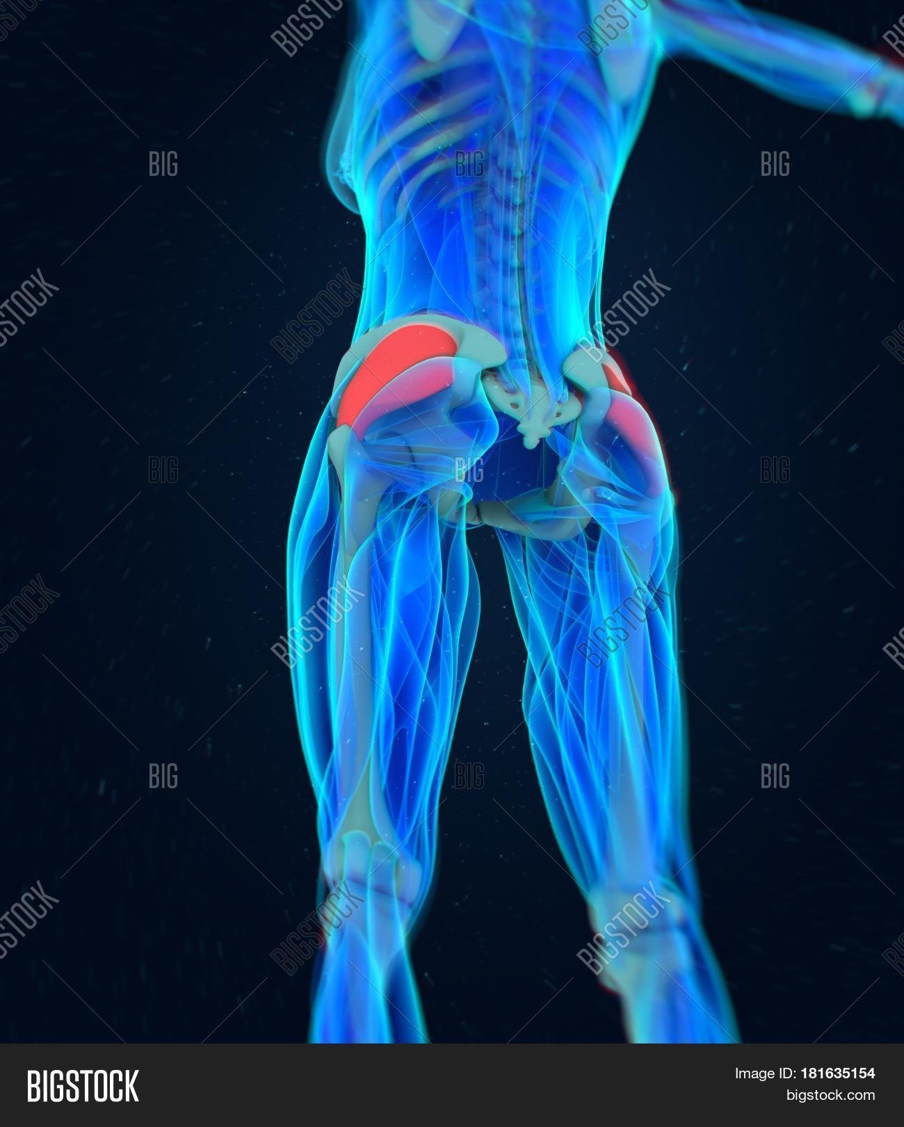
Female hip anatomy. Muscle and tendon anatomy of the hip adductors gluteal muscles or buttocks hamstring muscles femoral muscle quadrices. Iliopsoas muscle a hip flexor muscle that attaches to the upper thigh bone. The muscle originates from the body of the pubis and attaches to the pectineal line and proximal part of the linea aspera of femur.
Yet the hip joint is also one of our most flexible joints and allows a greater range of motion than all other joints in the body except for the shoulder. The anatomy of the pelvis varies depending on whether you are male or female. Rectus femoris muscle one of the quadriceps muscles on the front of your thigh.
A round cup shaped structure on the os coxa known as the acetabulum. Muscles play an important role in the health and well being. Since broad hips facilitate child birth and also serve as an anatomical cue of sexual maturity they have been seen as an attractive trait for women for thousands of years.
The female hips have long been associated with both fertility and general expression of sexuality. Together they form the part of the pelvis called the pelvic girdle. The testicles and scrotum are also important male structures.
The male pelvic organs include the penis and various glands and ducts. It is enervated by the obturator nerve. Use the mouse scroll wheel to move the images up and down alternatively use the tiny arrows on both side of the image to move the images on both side of the image to move the images.
The female pelvic bones are typically larger and broader than a males. This mri hip joint axial cross sectional anatomy tool is absolutely free to use. The male urethra and the penis the male urethra is a muscular tube that runs through the prostate perineal membrane.
The hip joint is a ball and socket type joint and is formed where the thigh bone femur meets the pelvis. Some of the other muscles in the hip are. There are two hip bones one on the left side of the body and the other on the right.
This is so a baby can pass through the pubic outlet the circular hole in the middle of the pelvic bones during childbirth. Captioned anatomical structures of the gluteal area buttocks ligaments. The hip joint is a ball and socket synovial joint formed between the os coxa hip bone and the femur.
48 adductor longus muscle this muscle is the most anterior of the adductor group of muscles in the thigh.
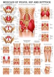 Pelvic Floor Musculature Laminated Anatomy Chart Amazon
 Female Character Production Guide Anatomy Tutorial Human
 Anatomy Hip Stock Illustrations 6 500 Anatomy Hip Stock
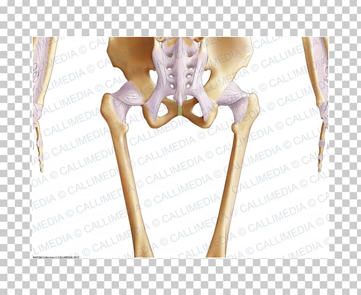 The Female Pelvis Anatomy Exercises Bone Hip Finger Png
 Artstation Female Pelvis Human Anatomy 3d Model Dean Kezan
 Stock Illustration The Female Leg Bones Clip Art
 Gluteus Medius Female Image Photo Free Trial Bigstock

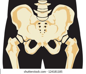 Royalty Free Female Hip Bone Stock Images Photos Vectors
 Hip Human Anatomy Inflammation Articular Joint Pain Stock
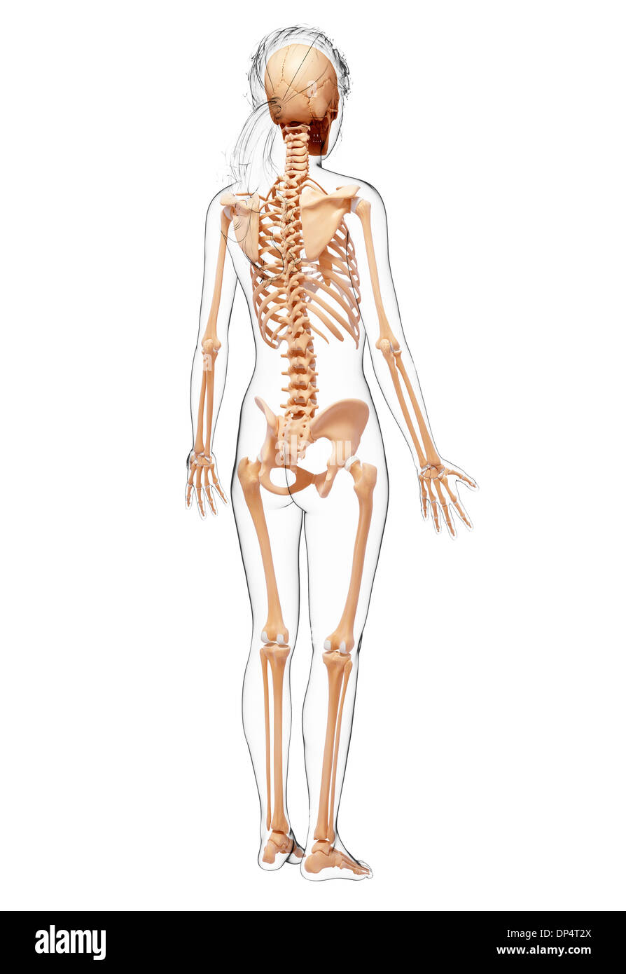 Female Hip Bone Cut Out Stock Images Pictures Alamy
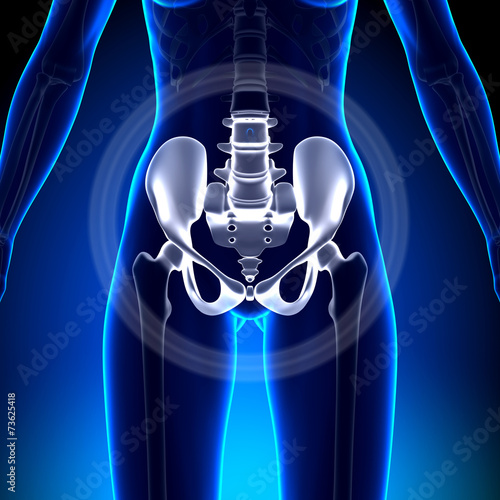 Female Hip Sacrum Pubis Ischium Ilium Anatomy
Xray Anatomy Of The Hip Review Xray Anatomy Of The Hip
 Female Hips Anatomy Stock Photos Page 1 Masterfile
 77 Best Hip Anatomy Images In 2019 Anatomy Anatomy
Mri Anatomy Of The Hip Review Mri Anatomy Of The Hip
 Pin By Deedee Austin On Study Reproductive System Female
 Anatomy Of Female Hips And Pelvic Bones Hardcover Journal
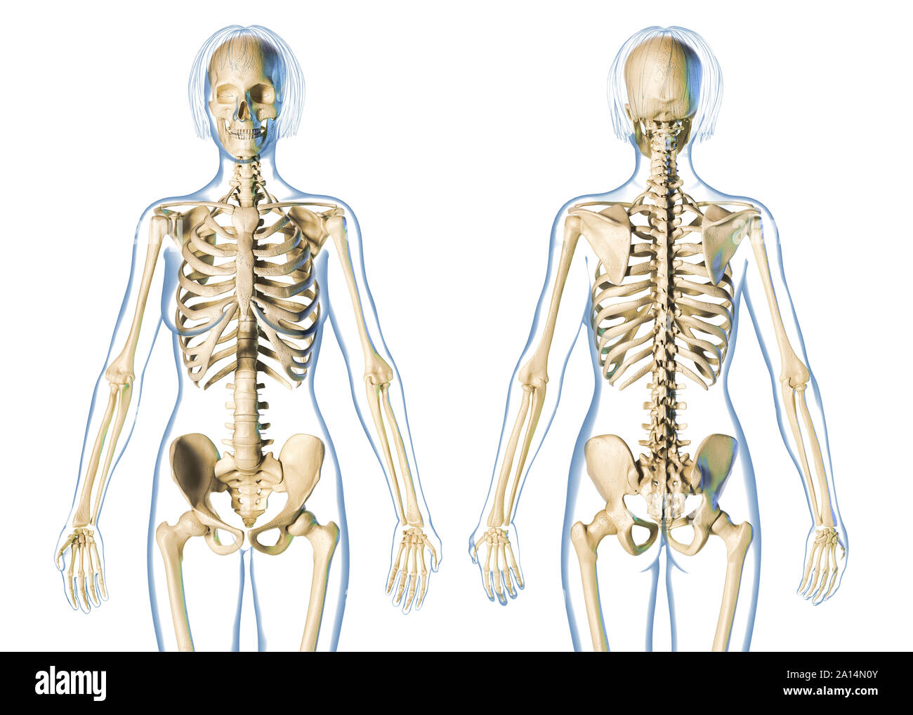 Female Hip Bone Stock Photos Female Hip Bone Stock Images
 Beautiful Hip Anatomy Art Fine Art America
:background_color(FFFFFF):format(jpeg)/images/library/11030/Hip_and_thigh_1.png) Hip And Thigh Bones Joints Muscles Kenhub
Hip And Thigh Bones Joints Muscles Kenhub
 The Most Popular Bodybuilding Message Boards In 2019

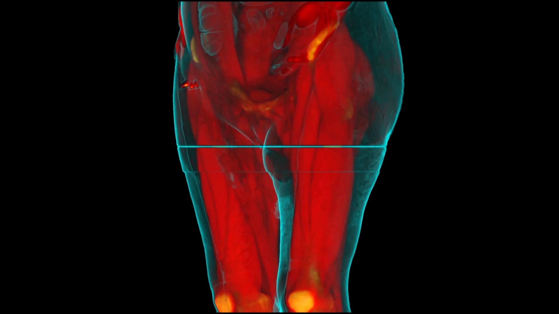

Belum ada Komentar untuk "Female Hip Anatomy"
Posting Komentar