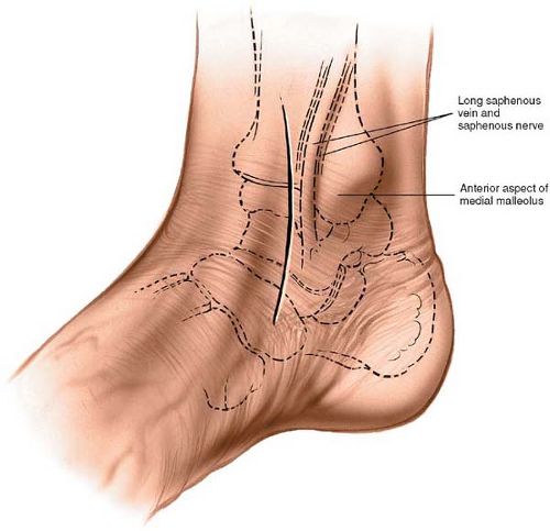
Medial Malleolus Anatomy
We think this is the most useful anatomy picture that you need. The lateral malleolus is the prominence on the outer side of ankle formed by the lower end of the fibula.
 This Trial Exhibit Depicts A Bimalleolar Right Ankle
The medial surface of this process is convex and subcutaneous.

Medial malleolus anatomy. This image added by admin. The tendon sheath of the posterior tibial muscle covers the posterior and middle part of the deltoid ligament in much the same way as the peroneal tendon sheath is associated with the calcaneofibular ligament on the lateral side. The posterior malleolus felt on the back of your ankle is also part of the tibias base.
The medial malleolus felt on the inside of your ankle is part of the tibias base. You can click the image to magnify if you cannot see clearly. Its lateral or articular surface is smooth and slightly concave and articulates with the talus.
It is attached above to the apex and anterior and posterior borders of the medial malleolus. Its posterior border presents a broad groove the malleolar sulcus directed. The medial surface of the tibia is prolonged downward to form a strong pyramidal process flattened from without inwardthe medial malleolus.
Mnemonics that can be used to remember the anatomy of the ankle tendons from anterior to posterior as they pass posteriorly to the medial malleolus under the flexor retinaculum in the tarsal tunnel include. Tom dick and very nervous harry. Its anterior border is rough for the attachment of the anterior fibers of the deltoid ligament of the ankle joint.
The lateral malleolus felt on the outside. Orthopaedicsone the orthopaedic knowledge networkcreated may 02 2010 1259. Tom dick and harry.
The medial malleolus is the prominence on the inner side of the ankle formed by the lower end of the tibia. The deltoid ligament or the medial ligament of talocrural joint is a strong flat and triangular band. The medial aspect of the distal extremity forms the medial malleolus.
Similar to the posterior talofibular ligament the mcl is a multifascicular ligament originating from the medial malleolus to insert in the talus calcaneus and navicular bone. The deltoid ligament is composed of superficial and deep components. Also called internal malleolus.
The medial malleolus surface anatomyorthopaedicsone articlesin. The bony bumps or protrusions seen and felt on the ankle have their own names. The rounded process of the tibia forming the internal surface of the ankle joint.
A medial process from the distal tibia which with the lateral malleolus forms a mortise into which the talus articulates. 484 the right tibia posterior view 821 the smooth posterior surface of the shaft of the tibia is marked by a prominent ridge the soleal line and a large oblong foramen the nutrient foramen.
 Anterior And Posterior Approaches To The Medial Malleolus
 Medial Malleolus An Overview Sciencedirect Topics
 5 Kinds Of Medial Malleolus Ankle Fractures
 Medial Ankle Ligament Physiopedia
 Pdf Functional Outcome Of Bimalleolar Ankle Fractures
 Ankle Lower Leg Anatomy Foot Ankle Lower Leg
 Ankle Fracture Of The Right Medial Malleolus Fracture
 Medial Malleolus Fracture Fractured Medial Malleolus
 The Tibia Proximal Shaft Distal Teachmeanatomy
 The Radiology Assistant Ankle Mri Examination
 Medial Malleolus Fracture And Broken Ankle Treatment
 Anatomy Ankle Joint Clinicals Medicine Tr051 Studocu
 The Radiology Assistant Ankle Fracture Mechanism And
 Image Result For Tenderness Posterior To The Medial
 Ankle Joint An Overview Sciencedirect Topics
 Anatomy Physiology Illustration
 Subcutaneous Bursa Of Medial Malleolus
 Ankle Anterior Approach Approaches Orthobullets
 Figure Tibia Fibula Fibular Notch Lateral
 30 September 2012 Gymnastics Injuries
 The Ankle Joint Articulations Movements Teachmeanatomy
 In A Comminuted Fracture Of The Fibula And A Medial
 Ultrasound Guided Saphenous Adductor Canal Block Nysora


Belum ada Komentar untuk "Medial Malleolus Anatomy"
Posting Komentar