Anatomy Of Diaphragm
Motor innervation of the diaphragm comes from the phrenic. The diaphragm is the main muscle of respiration and it separates the thorax from the abdomen and pelvis.
 Anatomy Of The Normal Diaphragm Semantic Scholar
It represents the floor of the thoracic cavity and the ceiling of the abdominal cavity.
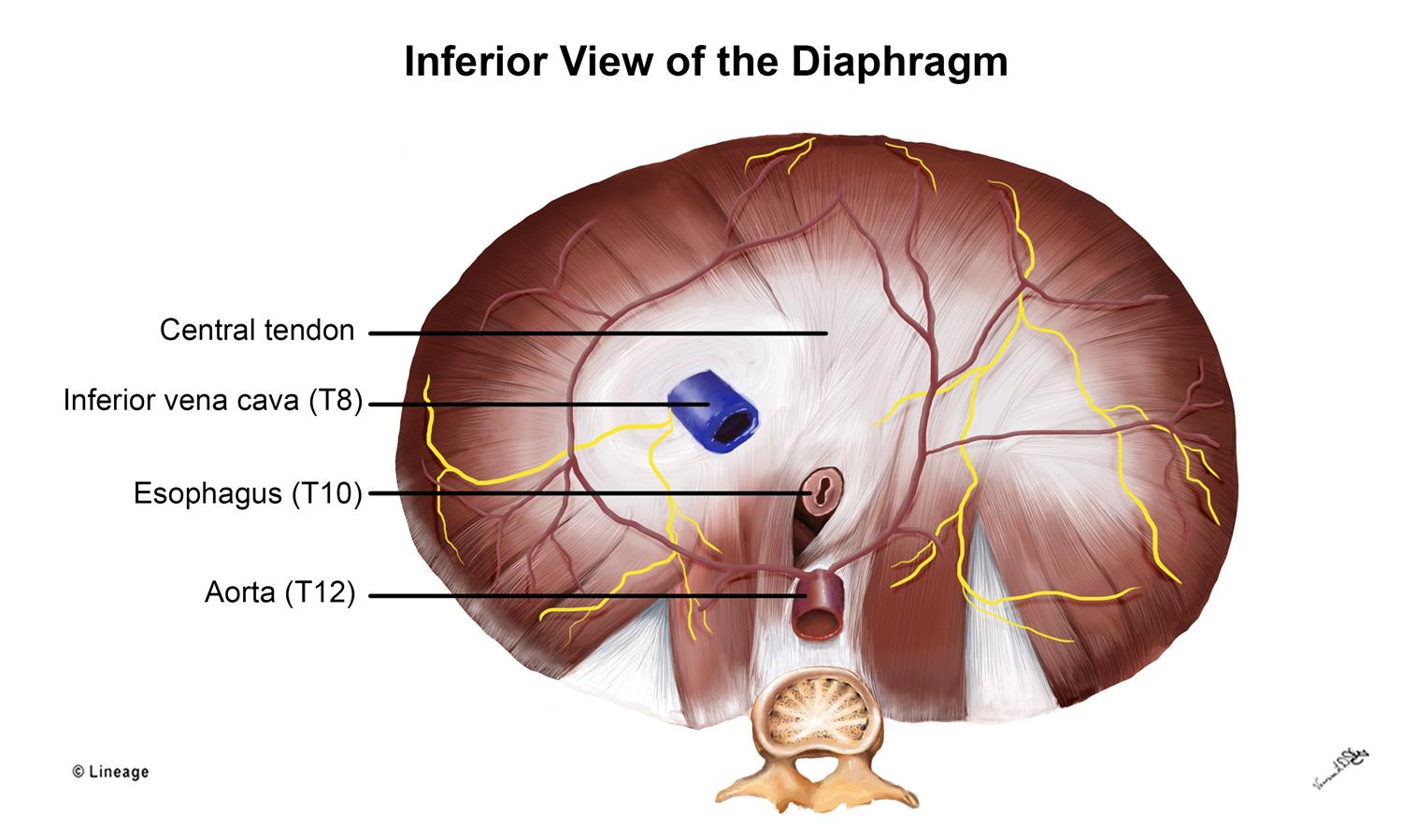
Anatomy of diaphragm. The diaphragm is a musculotendinous structure with a peripheral attachment. Structure anatomy of the diaphragm. Through which the esophagus passes.
It contracts and flattens when you inhale. Oesophagus 10 letters passes through the diaphragm at t10. The diaphragm is a musculotendinous sheet.
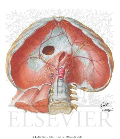
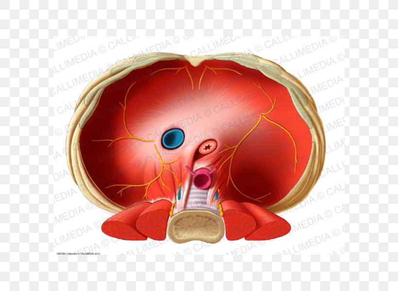
The diaphragm is pierced by many structures notably the esophagus aorta and inferior vena cava and is occasionally subject to herniation rupture. Vena cava 8 letters passes through the diaphragm at t8. It acts as the floor of the thoracic cavity and the roof of the abdominal cavity.
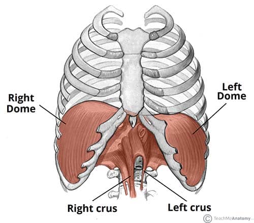
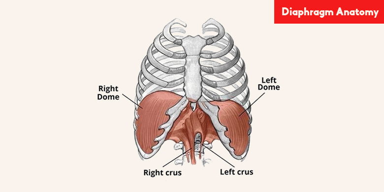
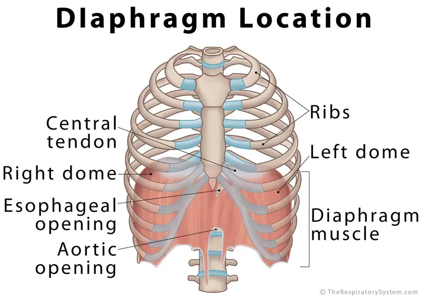
Also known as the thoracic diaphragm it serves as an important anatomical landmark that separates the thorax or chest from the abdomen. The thoracic spinal levels at which the three major structures pass through the diaphragm can be remembered by the number of letters contained in each structure. The right crus arises from the bodies of first three lumbar vertebrae and their intervertebral discs.
There are a number of bits of it worth talking about anatomically as things have to pass. There are 3 openings holes through the diaphragm. The diaphragm is one of the main muscles of respiration.
The diaphragm is the dome shaped sheet of muscle and tendon that serves as the main muscle of respiration and plays a vital role in the breathing process. Lateral to the crura on both sides the diaphragm arises from the medial and lateral arcuate ligaments. It acts as the floor of the thoracic cavity and the roof of the abdominal cavity.
The diaphragm is also important in expulsive actionseg coughing sneezing vomiting crying and expelling feces urine and in parturition the fetus. The diaphragm is located at the inferior most aspect of the ribcage filling the inferior thoracic aperture. Diaphragm anatomy and function the diaphragm is a thin skeletal muscle that sits at the base of the chest and separates the abdomen from the chest.
The diaphragm is a parachute shaped muscle that separates the chest from the abdomen.
 The Diaphragm Yogabody Anatomy Kinesiology And Asana
The Diaphragm Actions Innervation Teachmeanatomy
 Diaphragm And Posterior Abdominal Wall Human Anatomy 1
 Ventraal Organ Abdomen Anatomy Human Body Urogenital

 Anatomy Of The Normal Diaphragm Semantic Scholar
 Diaphragm Ajith Sominanda Department Of Anatomy Faculty Of
 Anatomy Of The Normal Diaphragm Semantic Scholar
 Diaphragm Anatomy 19th C Illustration Stock Image C029
 Diaphragm Abdominal Surface
![]() Diaphragm Muscle Anatomy Innervation And Function Kenhub
Diaphragm Muscle Anatomy Innervation And Function Kenhub
 What Is Diaphragm Amazing Facts About Diaphragm Function
 Thoracic Diaphragm Inferior Vena Cava Human Anatomy
 Diaphragm Respiratory Medbullets Step 1
 Diaphragm Radiology Key
 Diaphragm Muscle Images Stock Photos Vectors Shutterstock
 Yoga Anatomy 6 Reasons Why The Diaphragm May Be The Coolest
 Diaphragm Definition Function Location Britannica
 Diaphragm Definition Location Anatomy Function Diagram
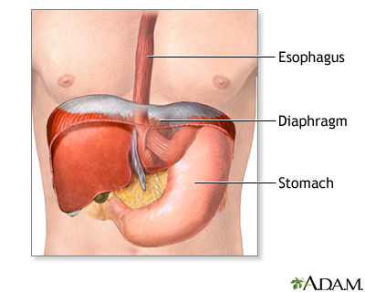 Hiatal Hernia Repair Series Normal Anatomy Medlineplus
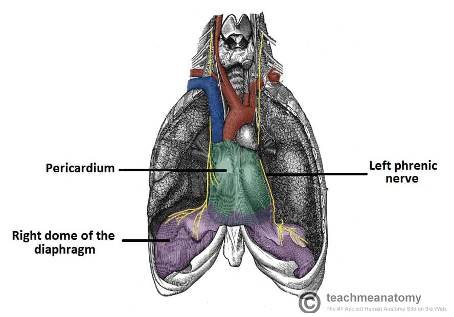 The Diaphragm Actions Innervation Teachmeanatomy
 Diaphragm An Overview Sciencedirect Topics
 Human Diaphragm Anatomy
 Instant Anatomy Diagram
 Image Result For Diaphragm Anatomy Thoracic Duct Xiphoid
![]() Human Diaphragm Anatomy Drawing Of A Normal Human Diaphragm
Human Diaphragm Anatomy Drawing Of A Normal Human Diaphragm
 Left Thorax Anatomy And Right Attachment Points Of The
 Amazon Com Anatomy Diaphragm Abdominal View Print Sra3
Belum ada Komentar untuk "Anatomy Of Diaphragm"
Posting Komentar