Dorsal Wrist Anatomy
We think this is the most useful anatomy picture that you need. These two terms used in anatomy and embryology refer to back dorsal and front or belly ventral of an organism.
Mri Of The Extensor Tendons Of The Wrist
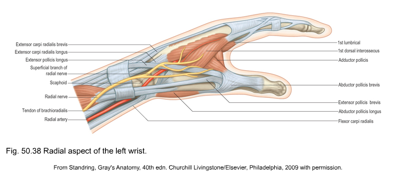
The abductor pollicis longus apl and extensor pollicis brevis epb.

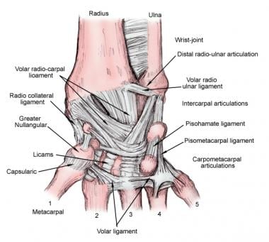
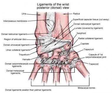
Dorsal wrist anatomy. Reinforced by the floor and septa of the fibrous tunnels for the six dorsal compartments see below and have a z shaped configuration. A series of ligaments that extend transversely across the palmar front or volar surface of the wrist connecting the carpals to one another and the carpals to the radiusulna. Dorsal ligaments palmar intercarpal ligaments.
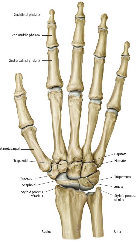
The dorsal from latin dorsum meaning back surface of an organism refers to the back or upper side of an organism. Bones of the wrist palmar view. It is actually a collection of multiple bones and joints.
Dorsal wrist the hand is pronated. The distal radioulnar joint is a pivot joint located between the bones of the forearm the radius and ulna. The wrist is a complex joint that bridges the hand to the forearm.
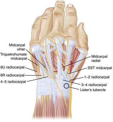
The dorsal wrist ligaments are comparatively thin. Listers tubercle serves as a good landmark between compartments 2 and 3. The wrist also contains several soft tissues such as ligaments tendons nerves and blood vessels.
The fibres of the dorsal radiocarpal ligaments are aligned more or less in the same axis as the forearm those of. During its examination the patients hand should rest freely or if a good access cannot be obtained it should be raised from the radial side. We think this is the most useful anatomy picture that you need.
These soft tissues function with the wrists bones and joints to provide movement sensation and nourishment to the hand. A transverse sweep across the dorsal wrist is sometimes helpful identifying the extensor compartments 1 6. Formed by the head of the ulna and the ulnar notch of the radius this joint is separated from the radiocarpal joint by an articular disk lying between the radius and the styloid process of the ulna.
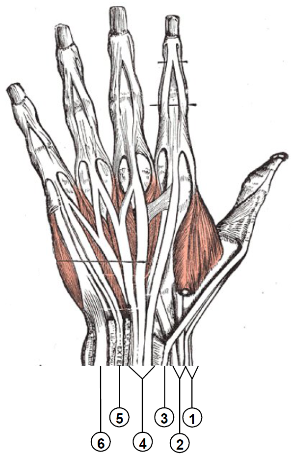
If talking about the skull the dorsal side is the top. The first compartment of extensor tendons is localized on the radial dorsal surface of the wrist and includes the tendons of. The bones comprising the wrist include the distal ends of the radius and ulna 8 carpal bones and the proximal portions of the 5 metacarpal bones see the images below.
 Wrist Hand Atlas Of Anatomy
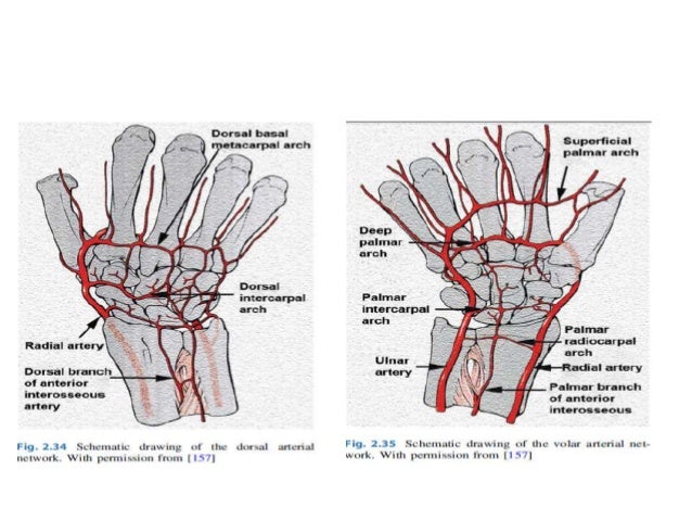
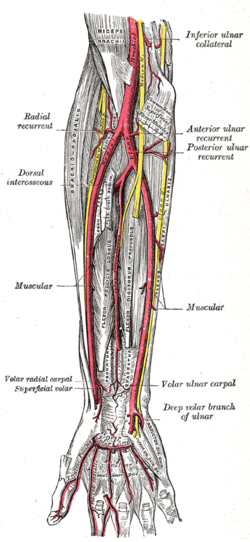 Radial Artery Wikipedia
 Conditions Affecting Dorsal Wrist Compartments Everything
![]() Wrist Anatomy Wrist Bones And Movements Kenhub
Wrist Anatomy Wrist Bones And Movements Kenhub
 Pin On Healthy Food
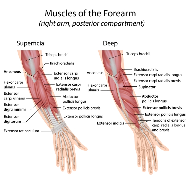 Muscles Of Hand And Wrist Bone And Spine
 Hamate Fractures The Hughston Clinic
 Lunate Dislocation Perilunate Dissociation Hand
 Ganglion Cyst Dorsal Wrist Fort Worth
 Wrist Joint Anatomy Overview Gross Anatomy Natural Variants

 Hand And Wrist Anatomy Pictures And Information
 Anatomy Of The Dorsal Aspect Of The Wrist Everything You Need To Know Dr Nabil Ebraheim
 Hand And Wrist Anatomical Poster Size 12wx17t Amazon Com
 Extensor Tendon Compartments Of The Wrist Wikipedia
 Wrist Joint Anatomy Overview Gross Anatomy Natural Variants
 Hand And Wrist Anatomy Volar View And Dorsal View Diagram
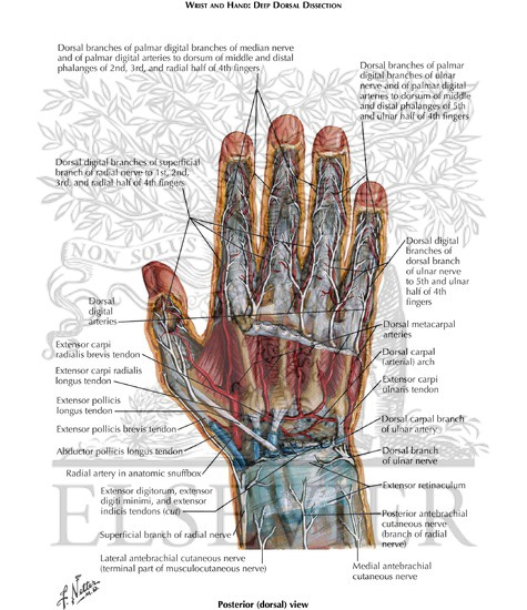 Wrist And Hand Deep Dorsal Dissection
 Evaluation And Diagnosis Of Wrist Pain A Case Based
 Wrist Hand Anatomy
 Wrist Block Landmarks And Nerve Stimulator Technique Nysora
 Wrist And Hand Ultrasound Radiology Key
 Wrist Arthroscopy Setup Anatomy And Portals
 Anatomy Of Wrist And Carpal Bones
 Dorsal Approach To The Wrist Approaches Orthobullets
 The Wrist Anatomy Flashcards Quizlet
Belum ada Komentar untuk "Dorsal Wrist Anatomy"
Posting Komentar