Knee Anatomy Ligaments
Another bone the patella kneecap is at the center of the knee. In the knee they give stability and strength to the knee joint as the bones and cartilage of the knee have very little stability on their own.
Two concave pads of cartilage strong flexible tissue called menisci minimize the friction created at the meeting of the ends of the tibia and femur.
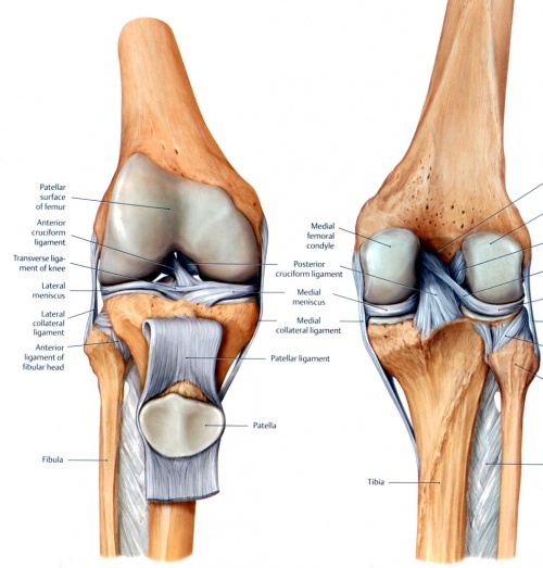
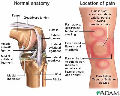
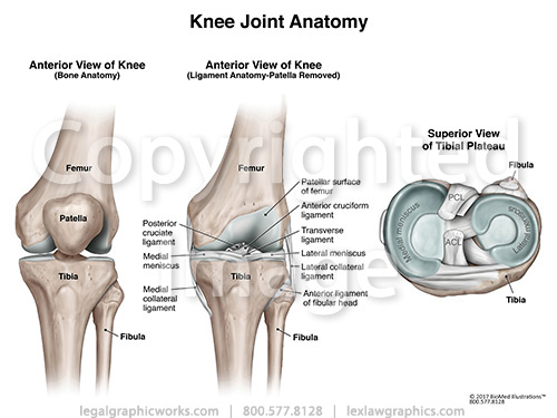
Knee anatomy ligaments. The medial collateral ligament mcl and lateral collateral ligament lcl are found on the sides of your knee. Two groups of muscles support the knee. The knee is a hinge joint that is responsible for weight bearing and movement.
Ligaments in the knee. One ligament is on each side of the knee joint. The anterior cruciate ligament and posterior cruciate ligament provide front and back anterior and.
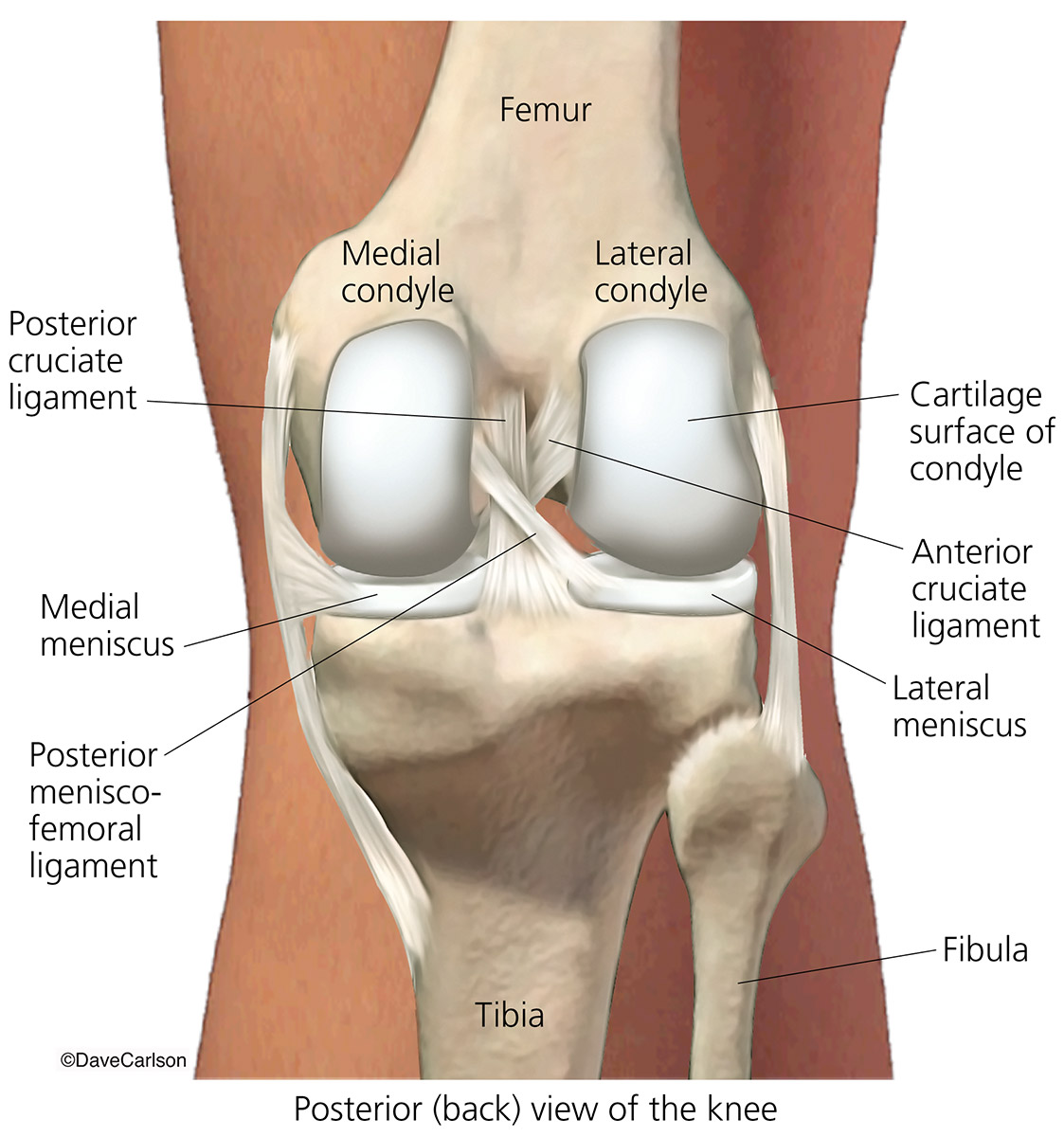
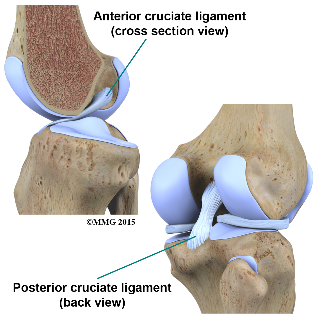
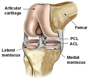
It consists of bones meniscus ligaments and tendons. Posterior cruciate ligament pcl which is located in the center of the knee and prevents excessive backward shifting of the knee. Knee ligament sprains or tears are a common sports injury.
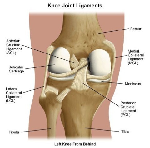
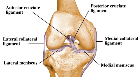
Ligaments of the knee. In knee joint anatomy they are the main stabilising structures of the knee acl pcl mcl and lcl preventing excessive movements and instability. Ligaments are strong tough bands that are not particularly flexible.
Ligaments join the knee bones and provide stability to the knee. The knee is designed to fulfill a number of functions. Your knee ligaments connect your thighbone to your lower leg bones.
Tendons connect the knee bones to the leg muscles that move the knee joint. The function of ligaments is to attach bones to bones and to help keep them stable. There are also several key ligaments a type of fibrous connective tissue that connect these bones.
There are four knee ligaments thick bands of tough tissue that serve to maintain the stability of the knee joint. On the sides of the knee are the medial collateral ligament mcl and the lateral collateral ligament lcl. The medial collateral ligament on the inner side and the lateral collateral ligament on the outer side.
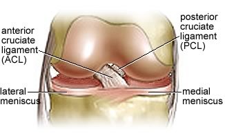
These two prevent sideways sliding of the knee joint ad also brace it against unusual movement. The anterior cruciate ligament prevents the femur from sliding backward on the tibia or the tibia sliding forward on the femur. Anterior cruciate ligament acl which is located in the center of the knee and prevents excessive forward movement of the tibia.
Ligaments are tough fibrous connective tissues which link bone to bone made of collagen. The most common ligament injuries are acl tears mcl tears. The knee includes four important ligaments all of which connect the femur to the tibia.
These are called the cruciate ligaments and consist of the anterior cruciate ligament and the posterior cruciate ligament. Knee ligament impose limitations on the movement of the knee allowing it to concentrate forces of the muscles on extension and flexion.
 The Knee Anatomy Injuries Treatment And Rehabilitation
 Cicitop Anatomy Science Medical Knee Joint With Ligaments
 Pain Behind Knee Why It Hurts In Back Of Or Under Your Kneecap
 Knee Joint Back View Carlson Stock Art
 Popliteal Ligament An Overview Sciencedirect Topics
/188058334-crop-56aae7425f9b58b7d0091480.jpg) What Is Causing Your Knee Pain
What Is Causing Your Knee Pain
 Collateral Ligament Cl Injury Aftercare Medlineplus
 Medial Patellofemoral Ligament Mpfl Reconstruction
 Physical Therapy In Buffalo For Knee Anatomy
 Care Of Your Knee Following Anterior Cruciate Ligament Acl
 Ligament Injuries Treatment Rochester Pcl Acl Injuries
 Anatomy Of The Knee Baxter Regional Medical Center
 The Knee Anatomy Injury Care And Prevention Soccer
 Types Of Knee Ligaments Stanford Health Care
 Knee Bones And Ligaments Artwork Stock Image C016 7011
 The Knee Explained Article Nzihf
 Ligaments Of The Knee Knee Sports Orthobullets
 Anatomy Of The Knee For Dancers Dance Work Balance
 The Human Knee Joint S Anatomy With Visible Cruciate
Common Knee Injuries Orthoinfo Aaos
 Knee Injuries For Parents Nemours Kidshealth




Belum ada Komentar untuk "Knee Anatomy Ligaments"
Posting Komentar