Lung Anatomy Hilar
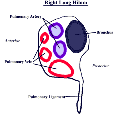
The hilum of the lung is a wedge shaped section in the central area of the lung that permits arteries veins nerves bronchi and other structures to enter and exit. Hilum of the lung.
 Lung Injuries Chapter 17 Atlas Of Surgical Techniques In
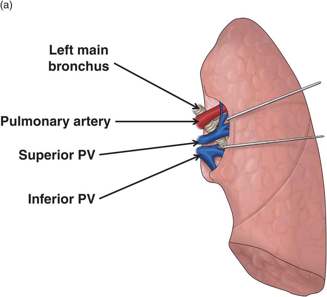
The pulmonary artery is the most superior structure within the left hilum.

Lung anatomy hilar. Lung roots lie opposite to t5 t7 vertebrae. Abnormalities in the hilum are usually noted on imaging. In the left hilum the left pulmonary artery occupies the upper part.
Both human lungs have a hilar region meaning both lungs have an area called the hilum. Gross anatomy left hilum. Tests to evaluate the hilum.
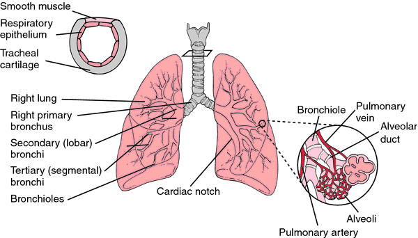
This concavity is deeper in the right lung due to the higher position of the right dome overlying the liver. Describe the root and hilum of lungs. The lung hilum where structures enter and leave the lung is located on this surface.
Both the right and the left lung have a hilum which lies roughly midway down. The base of the lung is formed by the diaphragmatic surface. Anatomy and abnormalities anatomy of the hilum.
Gross anatomy with the mediastinum at the hilum a circumscribed area where airways blood and. Lung root consists of the structures passing to and from the hilum of the lung to the mediastinum. It rests on the dome of the diaphragm and has a concave shape.
Hilum of the left lung. Below this is the left main bronchus. The left hilum is inferior and anterior to the aortic arch and thoracic aorta respectively.
The hilar region of. In human respiratory system. Structure of lung in lung to its apex is the hilum the point at which the bronchi pulmonary arteries and veins lymphatic vessels and nerves enter the lung.
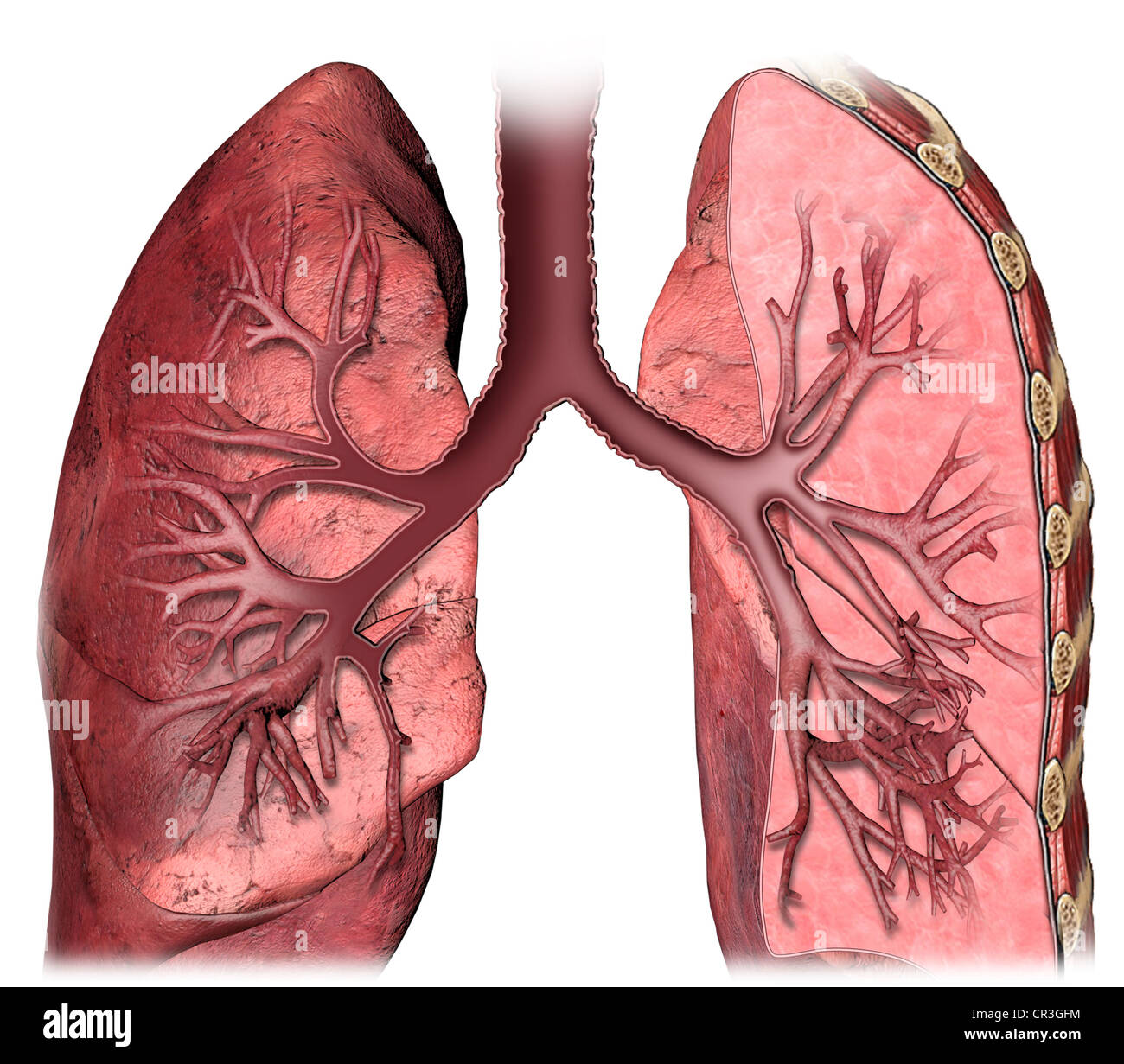
Hilum of lung a triangular depression where the structures which form the root of the lung enter and leave the viscus hilum of lymph node the portion of a lymph node where the efferent vessels exit. The lung hila or roots are found on the medial aspect of each lung. The left and right lung roots are similar but not identical.
Immediately below is the principal bronchus followed by the lower pulmonary veins. The structures of the lung root are embedded in the connective tissue and surrounded by extension. This region aids the lungs root in anchoring the lungs to.
 Lung Hilus Definition Of Lung Hilus By Medical Dictionary
Cadaveric Study Of Lung Anatomy A Surgical Overview
Hilum Of Lung Anatomy Unit 10 Diagram Quizlet
 Hilum Anatomy Britannica
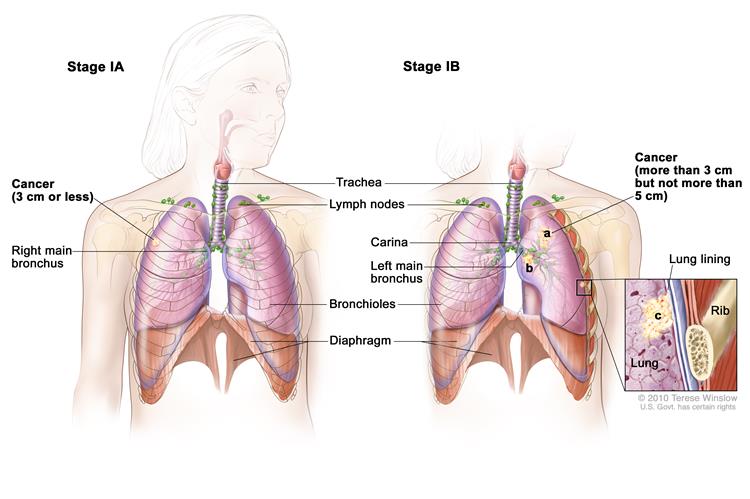
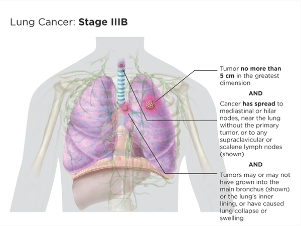 Lung Cancer Staging Lungevity Foundation
What Does Bilateral Hilar Congestion In A Chest X Ray
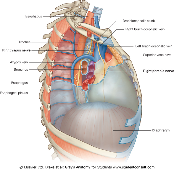
 Dissector Answers Superior Mediastinum Lungs
 The Lungs Position Structure Teachmeanatomy
 Lung Cancer Awareness Month
 Pulmonary Cavities Anatomy An Essential Textbook 1st Ed
 Help With A Simple Question I Cannot Find A Clear Answer To
 Non Small Cell Lung Cancer Staging Stages Of Lung Cancer
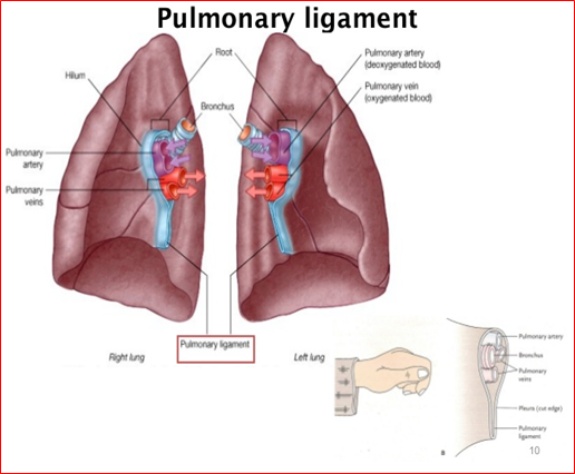
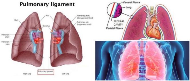 Pulmonary Ligament Pleurae Hilum Of The Lungs The Root Of

 Pulmonary Artery Wikipedia
 Pulmonary Vascular System And Pulmonary Hilum Sciencedirect
 Lung Cancer Physiopedia
 Tracheobronchial Sleeve Resection Technique Approach
 Lung Hilum Anatomy Stock Photo 48636664 Alamy
![]() Hilum Of The Lung Anatomy And Clinical Aspects Kenhub
Hilum Of The Lung Anatomy And Clinical Aspects Kenhub

 Left Lung Images Stock Photos Vectors Shutterstock
/iStock_000006469946_Large-56a5c5575f9b58b7d0de6a59.jpg) Hilum Of The Lung Definition Anatomy And Masses
Hilum Of The Lung Definition Anatomy And Masses
 Pleurae Pleural Cavity Pericardial Membrane Root Of Lung
 Anatomy Lung And Pleurae At King S College London Studyblue
 The Lungs Anatomy And Physiology Ii

Belum ada Komentar untuk "Lung Anatomy Hilar"
Posting Komentar