Transitional Lumbosacral Anatomy
This review will focus on the clinical significance of lstv disruptions in normal spine biomechanics imaging techniques diagnosis and treatment. In transitional lumbosacral vertebrae one of the vertebrae does not form as part of the lumbar or sacral area.
 Anteroposterior Lumbar Radiographs With Diagrams Overla Open I
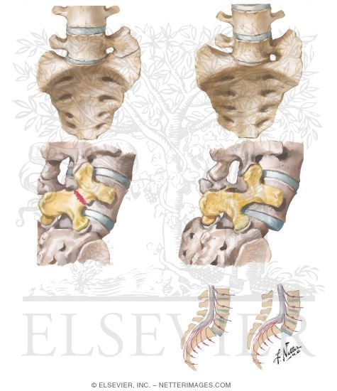
Facet joints even rudimentary and intervertebral disc between s1 and s2.
Transitional lumbosacral anatomy. These extra vertebrae usually occur at the lumbosacral juncture but can rarely also occur at the cervicothoracic frontier. Presence of six rib free lumbar type vertebrae which may have the following features squaring of highest sacral transitional vertebra. A transitional vertebra at the lumbosacral junction can cause arthritis disk changes or spinal cord compression.
Lumbosacral transitional vertebra lstv is a developmental spinal anomaly in which the lowest lumbar vertebra shows elongation of its transverse process and varying degrees of fusionfailure of segmentation from the sacrum. Lumbosacral transitional vertebrae lstvs are a congenital vertebral anomaly of the l5 s1 junction in the spine. It is as if this one vertebra cannot make up its mind whether to be become a lumbar vertebra or a sacral vertebra.
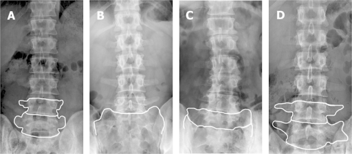
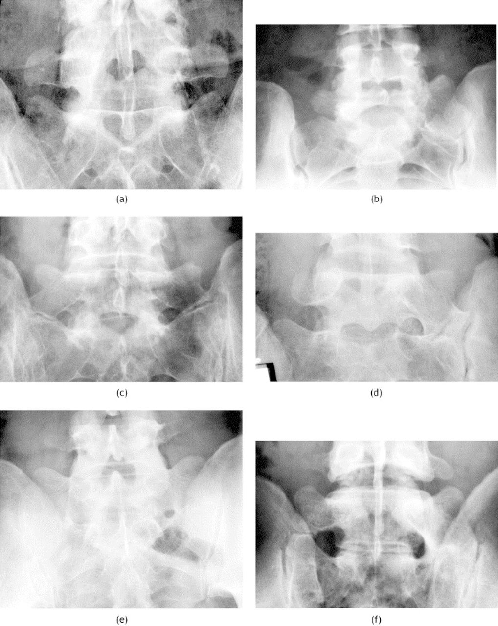
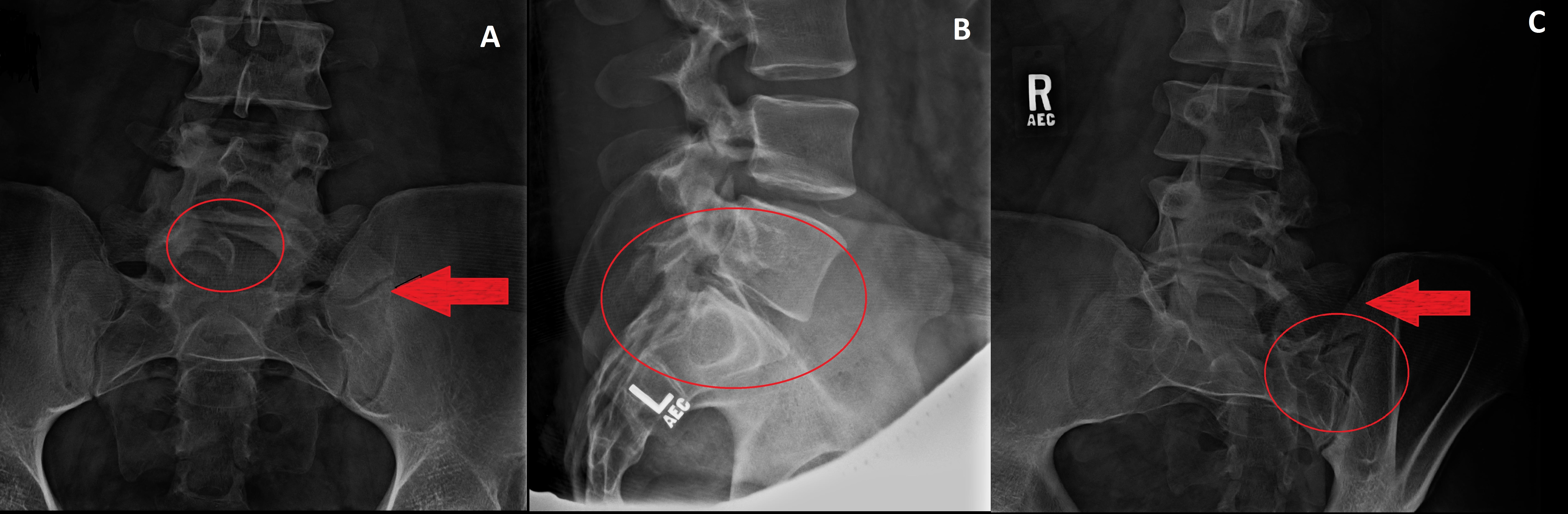
A transitional vertebra is an extra section of spinal bone that does not normally present in a typical anatomy. Lumbosacral transitional vertebrae lstv are increasingly recognized as a common anatomical variant associated with altered patterns of degenerative spine changes. Lumbosacral transitional vertebrae have been classically identified by using lateral and ferguson radiographs fig 1.
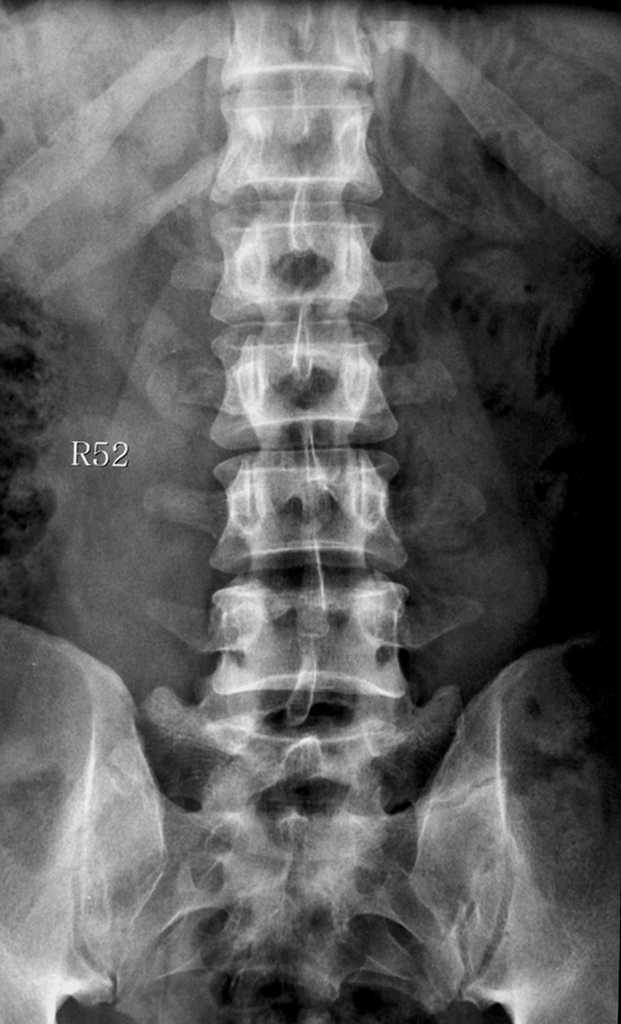
For instance the transverse process of the last cervical vertebra may resemble a rib. This is likely l5 with partial fusion of the right transverse processthere is suggestion of a pseudojoint which can be a source of pain. This alteration may contribute to incorrect identification of a vertebral segment leading to wrong level spine surgery and poor correlation with clinical symptoms.
The report says there is a transitional vertebra at the lumbosacral joint. It occurs at the cervicothoracic thoracolumbar or lumbosacral junction. The pedicles are intact and the sacroiliac joints are unremarkable.
The vertebral body height and the remainer of the disk spaces are well maintain ed. The human spine is made up of 33 bones called vertebrae with the spinal cord running through the middle. There is no fracture or spondylolisthesis.
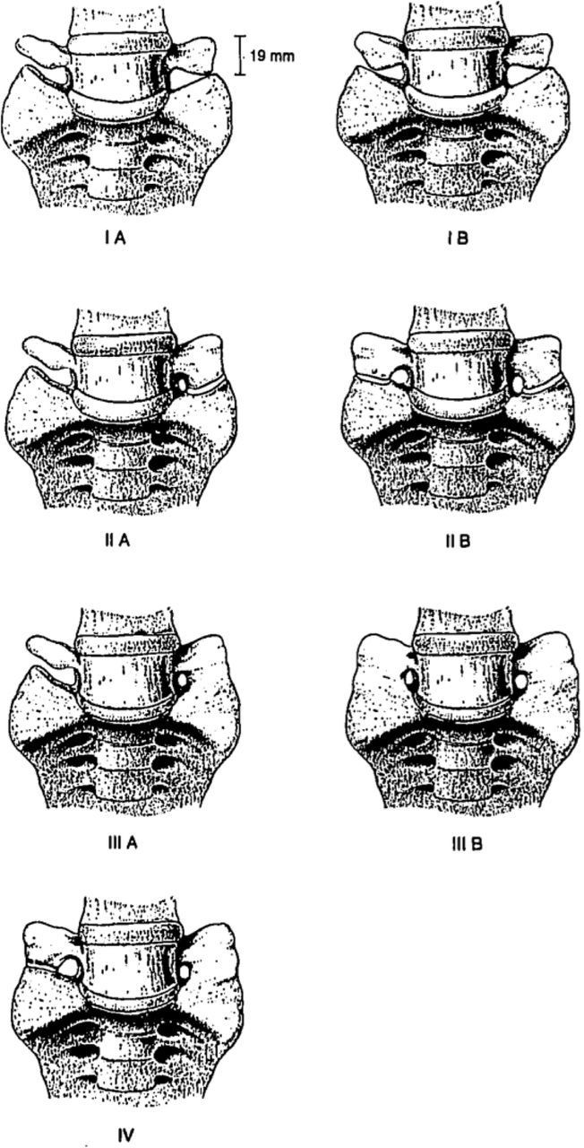
There is marked narrowing of the l5 s1 disk space as well. In 1984 castellvi et al described a radiographic classification system identifying 4 types of lstvs on the basis of morphologic characteristics fig 2. Transitional vertebrae are abnormally formed vertebral bones that display the characteristics of two different types of vertebrae.

 Lumbar Vertebrae An Overview Sciencedirect Topics

![]() Transitional Vertebrae
Transitional Vertebrae
 Sacralization Symptoms Causes Treatment And Outlook

 Lumbarisation And Sacralisation
![]() Transitional Vertebrae
Transitional Vertebrae
 Lumbar Spine Anatomy Overview Gross Anatomy Natural Variants
 Bertolotti Syndrome Report Of A Case Revista Colombiana
 A Review Of Symptomatic Lumbosacral Transitional Vertebrae


 A Review Of Lumbosacral Transitional Vertebrae And
 A Review Of Symptomatic Lumbosacral Transitional Vertebrae
 Lumbosacral Transitional Vertebra Castellvi Type Iib
 Lumbosacral Transitional Vertebrae Spondylolysis And
 Ecr 2016 C 0173 Lumbosacral Transitional Vertebrae Epos

 Incidence Rate Of Lumbosacral Transitional Vertebrae And
 Imaging Of Lumbosacral Transitional Vertebrae Sciencedirect

 Lumbar Spine Anatomy Spine Orthobullets

![]() A Ventrodorsal Radiograph Of A German Shepherd Dog With
A Ventrodorsal Radiograph Of A German Shepherd Dog With
 An Unwelcome Side Effect Transitional Vertebrae In Horses
 The Back Of The German Shepherd Dog The German Shepherd Dog
 Cureus Transitional Vertebra And Spina Bifida Occulta
Belum ada Komentar untuk "Transitional Lumbosacral Anatomy"
Posting Komentar