Venous Anatomy Leg
Anatomy physiology module provides a broad spectrum of adult male adult female and pediatric normal anatomy cases with varying body morphologies to maximize training efficacy. Often the artery and vein are located within the same vascular sheath so that the arterial pulsations aid the venous return.
 Venous Anatomy Sciencedirect
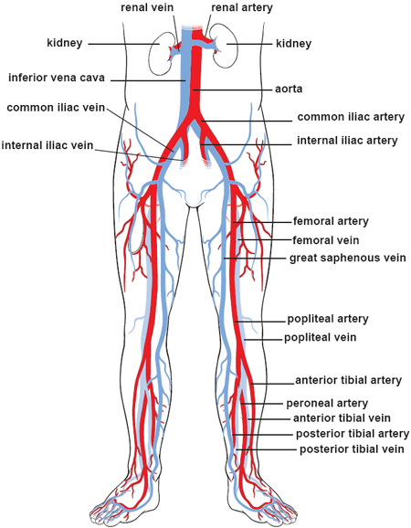
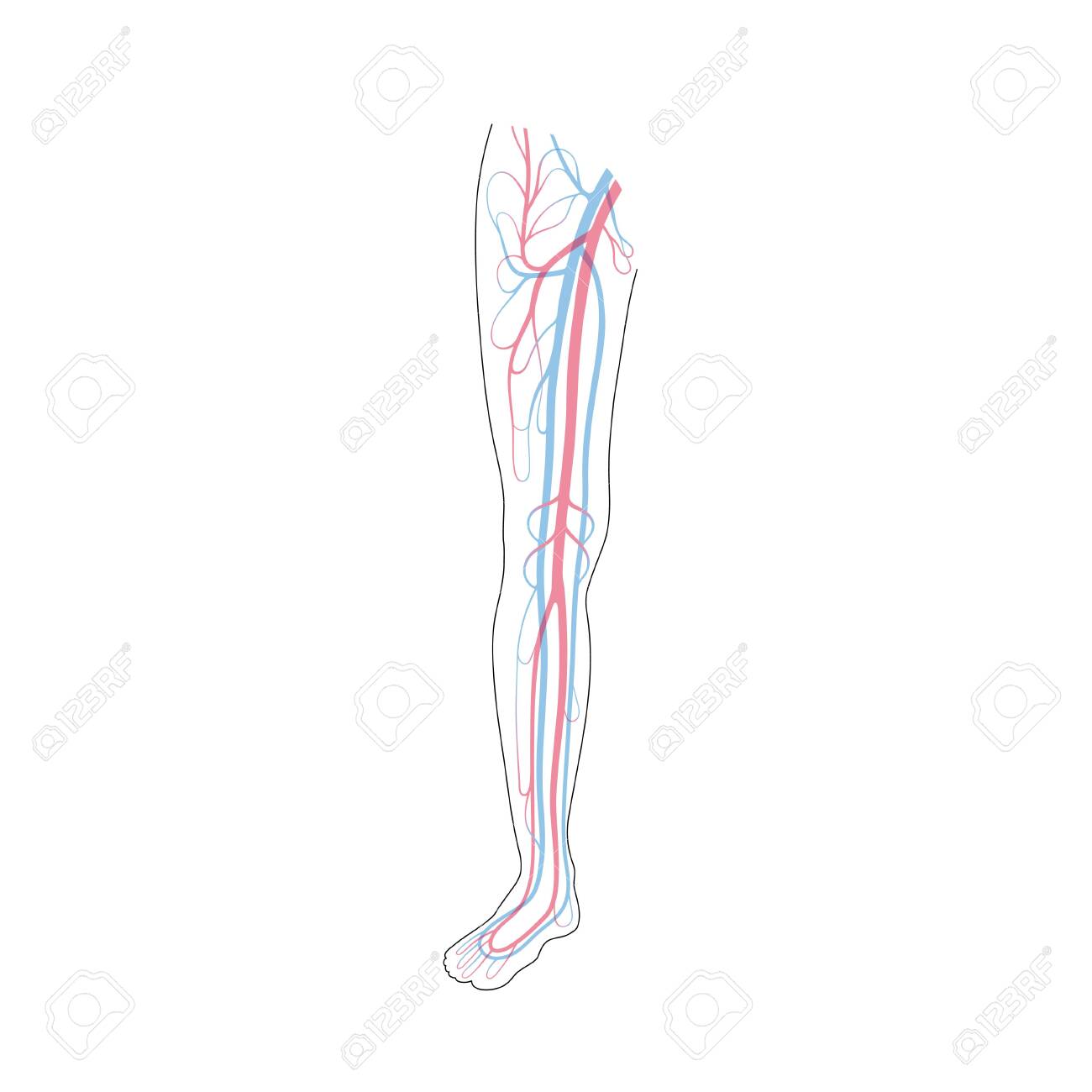
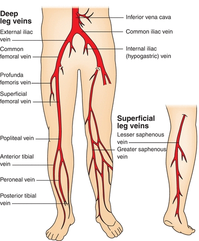
Anterior tibial vein which receives blood from the dorsal venous arch.

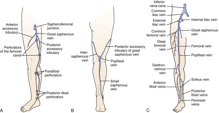
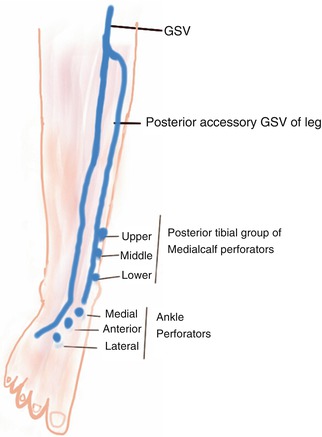
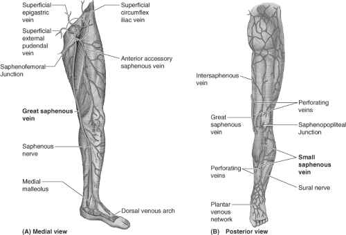
Venous anatomy leg. The foot and leg. The anterior accessory gsv of the leg drains the anterior aspect of the leg below the knee. This accounts for 95 of venous return.
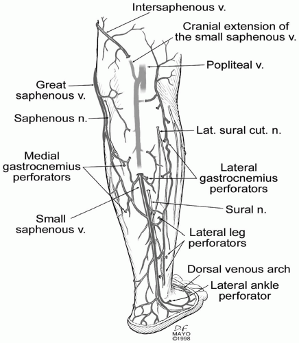
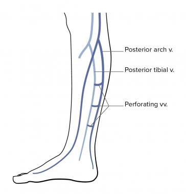
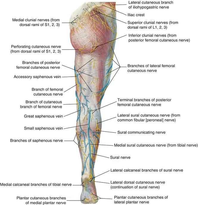
Posterior tibial vein and fibular vein also known as the peroneal vein which form from the medial and lateral plantar veins. The main venous structure of the foot is the dorsal venous arch which mostly drains into the superficial veins. The short saphenous vein has 7 13 valves and lies near the sural nerve within the leg.
Deep veins of the lower limb. The posterior accessory gsv of the leg leonardos vein or posterior arch vein is a common tributary it begins posterior to the medial malleolus ascends on the posteromedial aspect of the calf and joins the gsv distal to the knee see figure 28. A majority of the blood in the legs travels through the large major veins located in the limbs deep areas.
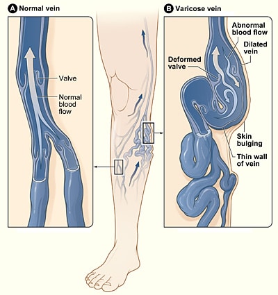
There are three main deep veins in the lower leg. It has many branches which communicate with the long saphenous vein. The rest is carried by the superficial veins which are the veins we see right underneath the skin and cause unsightly varicose veins.
On the dorsum of the foot deep veins drain into the short saphenous vein and within the leg it receives many cutaneous tributaries. Some veins from the arch penetrate deep into the leg forming the anterior tibial vein. Each individual hands on training case is accompanied by image window specific expert instruction and probe positioning guidance.
 Leg Venous Anatomy Physiology Module Sonosim
 Image Result For Lower Extremity Venous Anatomy Arteries
 Venous Physiology Clinical Gate
 Assessment And Management Of Older People With Venous Leg Ulcers

 Vein Care Specialist Olympia Tacoma Gig Harbor Wa
 Vein Services Biltmore Cardiology
 The Left Panel Shows The Anterior View Of Veins In The Legs
 Leg Knee Anatomy
 Drawing Of The Veins Of The Leg And The Calf Muscle Pump
 Lower Limb Venous Anatomy Thoracic Key
 Venous Disease Thoracic Key

 Imagenes Fotos De Stock Y Vectores Sobre Venous Anatomy

 Phlebology And Treatment Of Leg Veins Plastic Surgery Key
 Schematic View Of Venous Anatomy From Insightful Phlebology
 Which Three Venous Systems Are Affected By Chronic Venous
 Vector Isolated Illustration Of Human Arterial And Venous Circulatory
 Imagenes Fotos De Stock Y Vectores Sobre Veins Leg Anatomy
 The Venous System Of The Foot Anatomy Physiology And
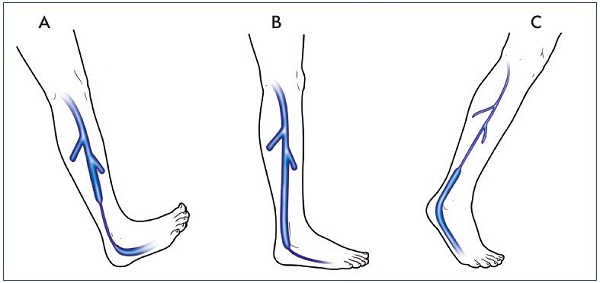
 Figure 3 From The Hemodynamics And Diagnosis Of Venous
 13101 01x Arteries And Veins Anatomy Exhibits
 Great Saphenous Vein Anatomy Pictures And Information
 Anatomy Springerlink
 Endovascular Treatment Of Varicose Veins Basicmedical Key
 Pdf Lower Extremity Venous Anatomy Semantic Scholar
 Lower Extremity Venous Anatomy Dallas Tx Venous System
 Varicose Veins Clinical Features Management
 Venous Disease And Lymphedema Management Nurse Key
 Venous Anatomy Sciencedirect
Belum ada Komentar untuk "Venous Anatomy Leg"
Posting Komentar