Zygoma Anatomy
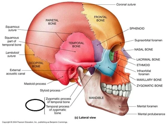
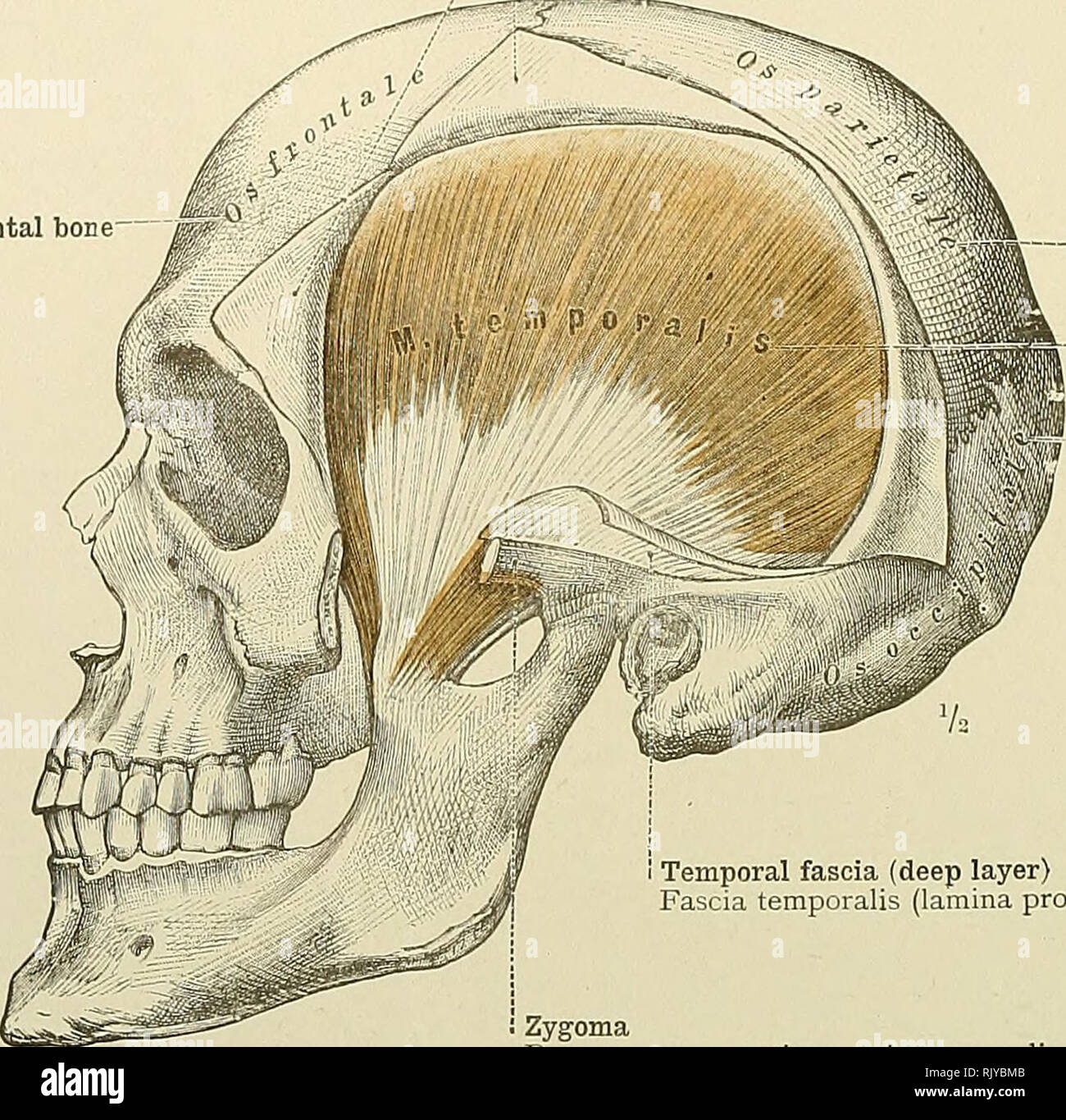
Another major chewing muscle the temporalis passes through the arch. Zygomatic bone also called cheekbone or malar bone diamond shaped bone below and lateral to the orbit or eye socket at the widest part of the cheek.
![]() Cunningham S Text Book Of Anatomy Anatomy Beanches Of The
Cunningham S Text Book Of Anatomy Anatomy Beanches Of The
The zygomatic bone is somewhat rectangular with portions that extend out near.

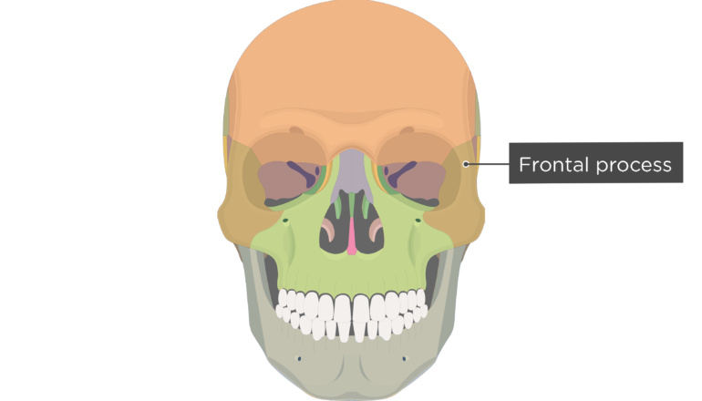
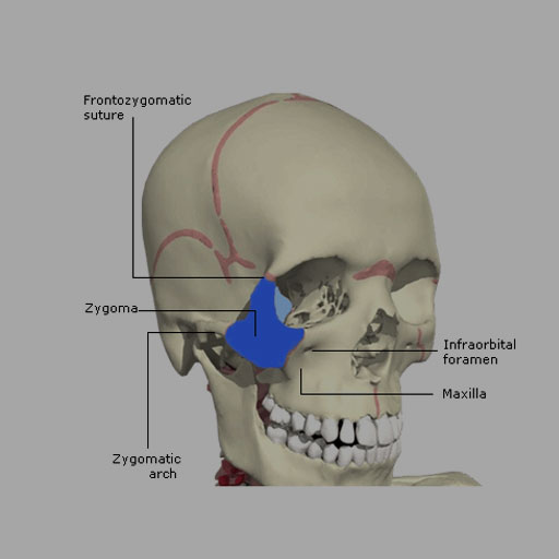
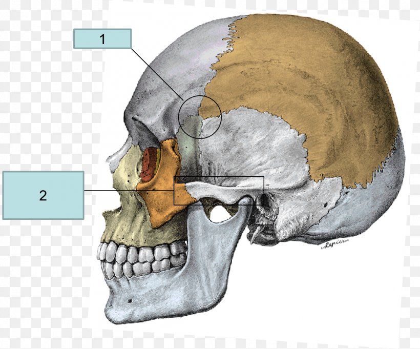
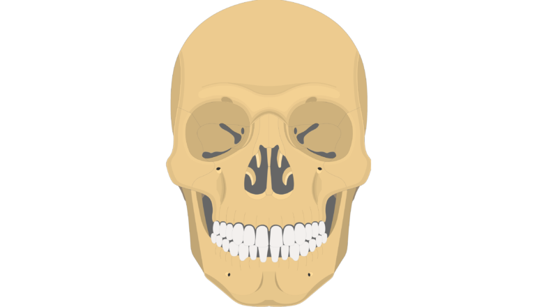
Zygoma anatomy. The zygoma also known as zygomatic bone or malar bone is an important facial bone which forms the prominence of the cheek. Zygomatic process of the maxillary bone articulated by the. Frontal bone via the frontozygomatic suture which creates the rounded form of the bony orbit.
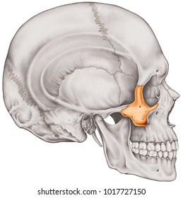
The masseter muscle important in chewing arises from the lower edge of the arch. It is situated at the upper and lateral part of the face and forms the prominence of the cheek part of the lateral wall and floor of the orbit and parts of the temporal fossa and the infratemporal fossa. The zygomatic bone is small and quadrangular and is situated at the upper and lateral part of the face.
Facial bone located below each eye socket anatomy. Zygomatic process of the temporal bone linked by the temporozygomatic suture. Introduction to temporal bone anatomy.
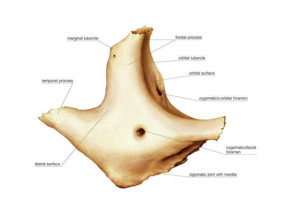
It adjoins the frontal bone at the outer edge of the orbit and the sphenoid and maxilla within the orbit. Gross anatomy zygoma has three surfaces five borders and two processes. The zygomatic bones gr zygoma yoke are two facial bones that form the cheeks and the lateral walls of the orbits.
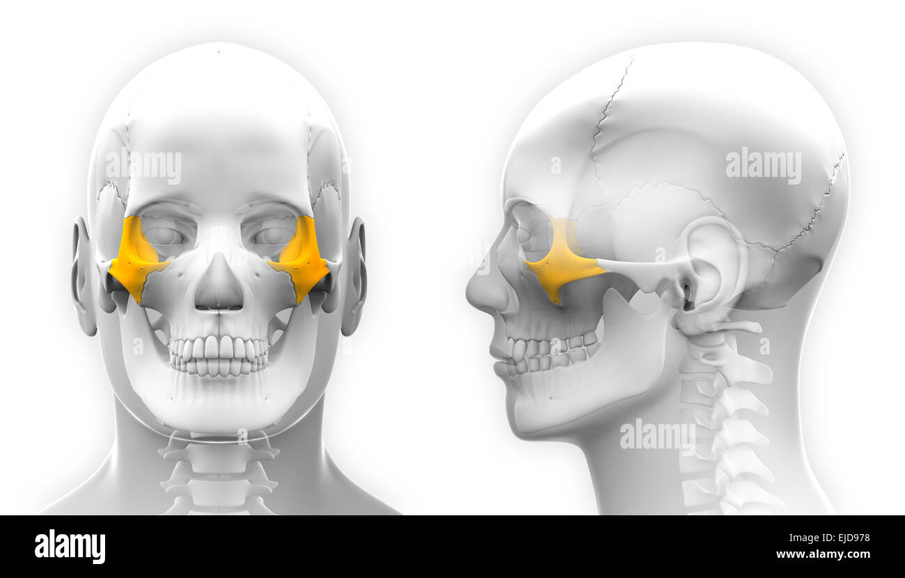
The most common condition associated with the zygomatic bone is. In the human skull the zygomatic bone cheekbone or malar bone is a paired irregular bone which articulates with the maxilla the temporal bone the sphenoid bone and the frontal bone. They are also commonly referred to a as the cheekbones or malar bones l mala the cheek.
Each zygomatic bone articulates with the temporal bone frontal bone maxilla and sphenoid bones. It is roughly quadrangular in shape. The zygomatic bone functions as a structure which joins the bones.
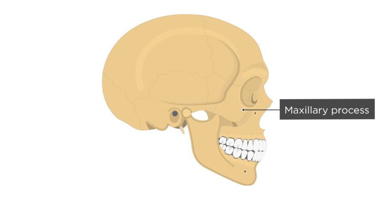
Four processes the frontosphenoidal orbital maxillary and temporal. Zygomatic arch bridge of bone extending from the temporal bone at the side of the head around to the maxilla upper jawbone in front and including the zygomatic cheek bone as a major portion. It forms the prominence of the cheek part of the lateral wall and floor of the orbit and parts of the temporal and infratemporal fossæ fig.
Several bones and joints surround the zygoma including the. It presents a malar and a temporal surface.
 Ce4rt X Ray Positioning Guide For Radiologic Techs
 The Cyclopaedia Of Anatomy And Physiology Anatomy
 Temporal Fossa Zygomatic Arch Ranzcrpart1 Wiki Fandom
 An Atlas Of Human Anatomy For Students And Physicians
 Zygomatic Bone Anatomy
 Zygomatic Bone

 Presurgical Examination Of Zygoma Anatomy Evaluated With
 Zygomatic And Nasal Injury Rcemlearning
 Ancestral Variations In The Shape And Size Of The Zygoma
 Superior Nasal Concha Zygomatic Bone Anatomy Temporal Bone
 Zygomatic Bone Wikipedia
 Benefits Of Zygomatic Implants In Patients With Severe
 Make Up For Special Effects Anatomy And Skeletal System Of
Zygomatic Process An Overview Sciencedirect Topics
 Zygomatic Bone
 Zygomatic Bone
 Zygomatic Bone Anatomy
 Midface Reduction Fixation Orif 4 Point Fixation
 Royalty Free Zygomatic Stock Images Photos Vectors
 Zygomatic Bone Stock Photos Zygomatic Bone Stock Images
 Zygoma Anatomy In 3d
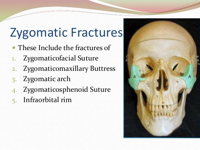
 32 Fractures Of The Zygoma Short Notes In Plastic Surgery
 Ii Osteology 5b 4 The Zygomatic Bone Gray Henry 1918
 Zygomatic Bone Anatomy
 Serdev Sutures In Middle Face Intechopen
 Rt 233 Skull Radiography Introducing Zygomatic Arches Ppt
 Zygomatic Process Of The Temporal Bone Zygomatic Arch
 Management Of Zygomatic Complex Fractures
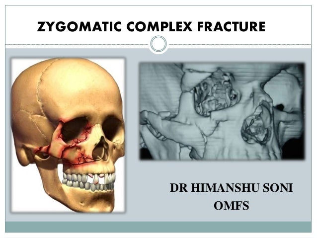 Zygomatic Complex Fracture Zmc
 Bones Of The Skull Zygomatic Bone Zygoma
Belum ada Komentar untuk "Zygoma Anatomy"
Posting Komentar