Brain Ct Scan Anatomy
Anatomy of the brain. Explore over 6700 anatomic structures and more than 670 000 translated medical labels.
 Introduction To Ct Head Approach And Principles Youtube
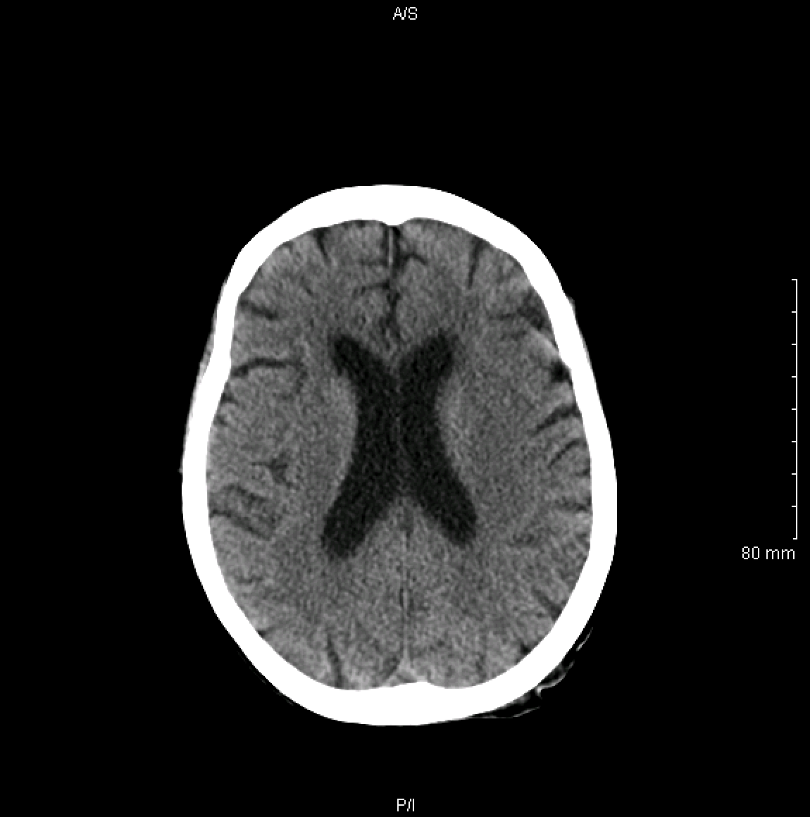
Ct scan provides a 3d display of the intracranial anatomy built up from a vertical series of transverse axial tomograms each tomogram represents a horizontal slice through the patients head.

Brain ct scan anatomy. This module is a comprehensive and affordable learning tool for medical students and residents and especially for neuroradiologists and radiation oncologists. This means that the right side of the brain is on the left side of the viewer. It is the most complete reference of human anatomy available on web ipad iphone and android devices.
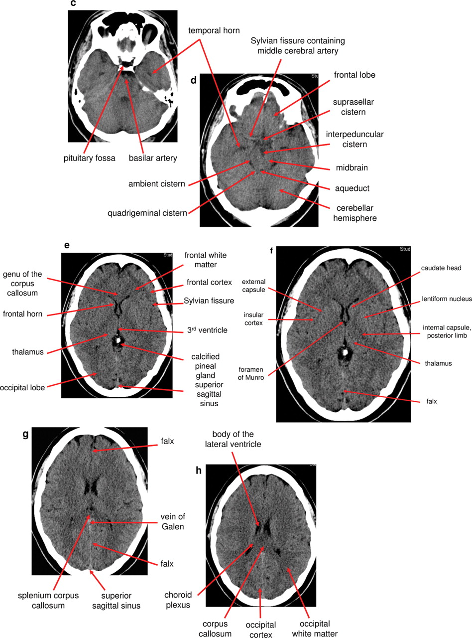
Dural venous sinuses veins arteries. Interactive anatomy atlas. This article lists a series of labeled imaging anatomy cases by system and modality.
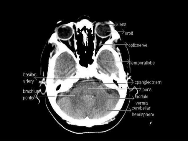
Ct brain image orientation. The scanner emits x rays towards the patient from a variety of angles and the detectors in the scanner measure the difference between the x rays that are absorbed by the body and x rays that are transmitted through the body. Head ct anatomy normal anatomy 1.
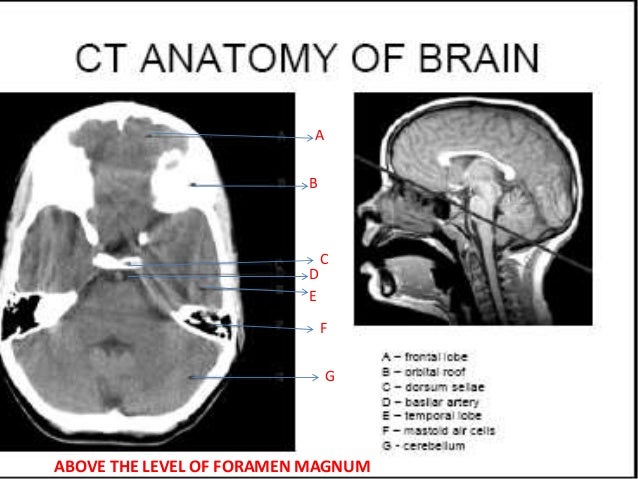
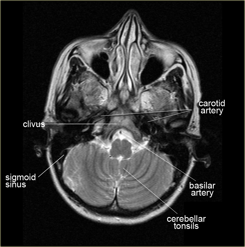
6 frontal bone 27 occipital bone 32 optic nerve 43 frontal sinus 45 sigmoid sinus 46 internal carotid artery. Available in 11. Ct mri radiographs anatomic diagrams and nuclear images.
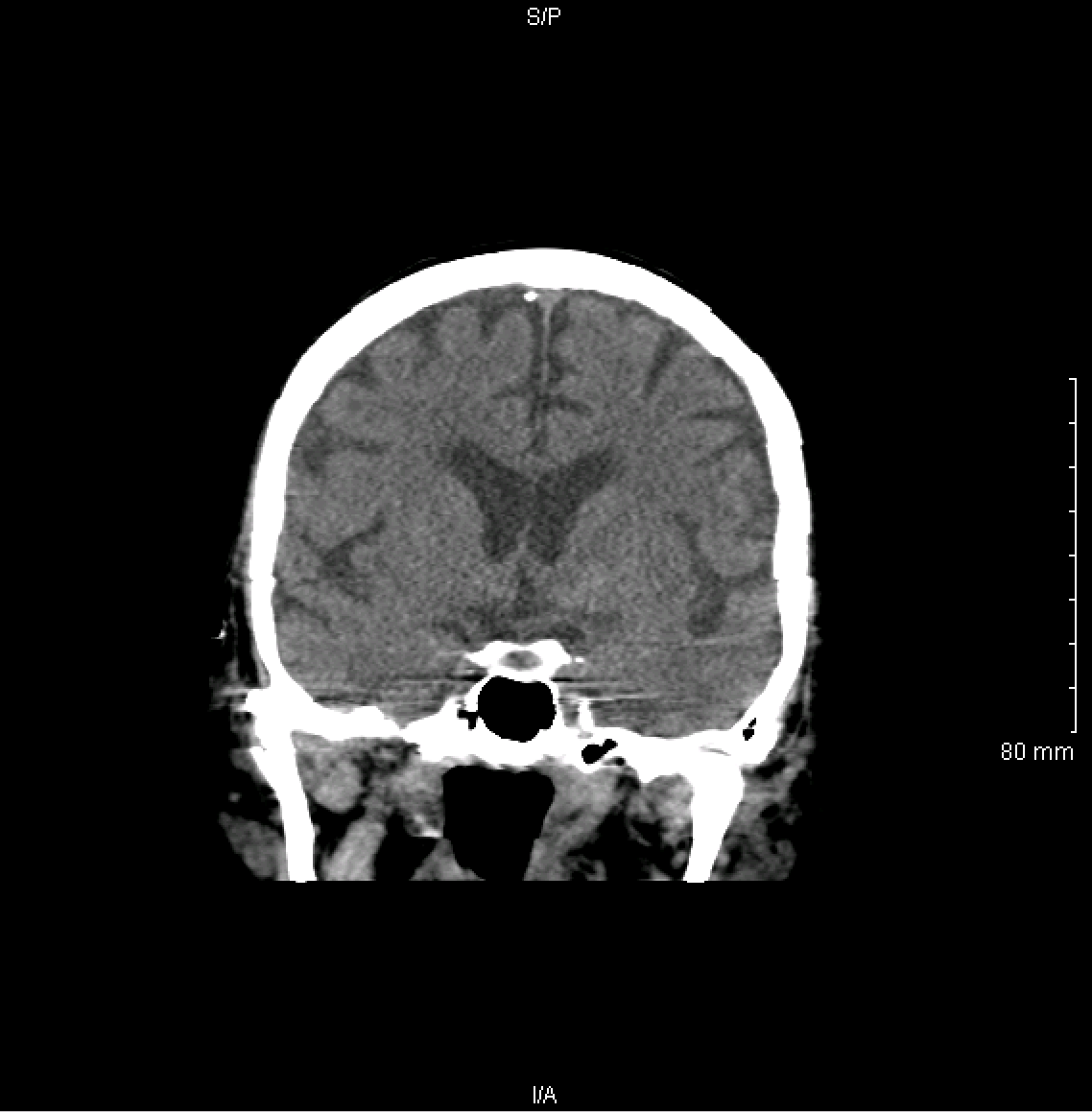
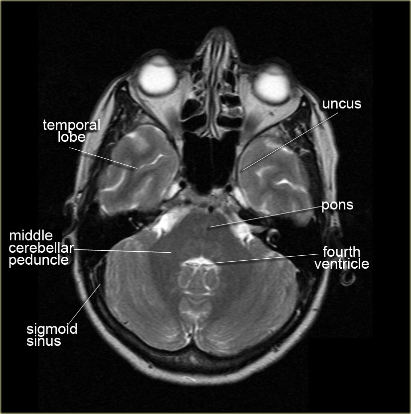
Anatomy of the head on a cranial ct scan. E anatomy is an award winning interactive atlas of human anatomy. Coronal brain ct.
Ct images of the brain are conventionally viewed from below as if looking up into the top of the head. Anatomy of the head on a cranial ct scan. Brain bones of cranium sinuses of the face.
Brain and face ct. Non contrast coronal ct head. Angiogram axial ct head.
Angiogram coronal ct head. Brain bones of cranium sinuses of the face. Non contrast axial ct head.
It provides access to an atlas and to images in axial planes allowing the user to learn and review neuroanatomy interactively. Non contrast sagittal ct head. Anatomy ct axial brain anatomy ct axial brain form no 1.
Anatomy ct axial brain form no 19. Ct scans are created using a series of x rays which are a form of radiation on the electromagnetic spectrum. How to view anatomical labels.
The anterior part of the head is at the top of the image. Learn ct scan learn the diagnosis of ct and methods of computed tomography.
 Ct Scans Interpretation Principles Basics Teachmeanatomy
 Normal Anatomy Of The Brain On Ct And Mri With A Few Normal
 Ct Scan Of The Head A Radiologist S Approach
 Head Ct
 Brain Imaging
 Normal Ct Brain
 Introduction To The Ct Brain
 Learn Ct Scan Anatomy Ct Axial Brain
 Basic Ct Anatomy Of The Brain
 Interpreting A Non Contrast Head Ct Scan Stepwards
 The Radiology Assistant Brain Anatomy
 How To Interpret An Unenhanced Ct Brain Scan Part 2
 Mri Anatomy Free Mri Axial Brain Anatomy
 Ct Brain Hemorrhage Startradiology
 Normal Head Ct
 Normal Head Ct Scan Anatomy Made Simple Neuroradiology
 How To Interpret An Unenhanced Ct Brain Scan Part 1 Basic
 Interpreting A Non Contrast Head Ct Scan Stepwards
 Ct Brain Hemorrhage Startradiology
 Ct Head Interpretation Radiology Geeky Medics
![]() The Ct Anatomy Tutor
The Ct Anatomy Tutor
 Confluence Mobil Tum Wiki
 Ct Anatomy
 Ct Scan Tips Protocols Ct Brain Anatomy
 Ct Head Axial Labeling Questions Radiology Case
 The Radiology Assistant Brain Anatomy
 Brain Imaging
 Axial View Of A Head Computed Tomography Ct Scan Of Pineal
 Crash Ctscan Enlargments Ct Brain Anatomy Radiology
 Interpreting A Non Contrast Head Ct Scan Stepwards
Introduction To Brain Surface Anatomy


Belum ada Komentar untuk "Brain Ct Scan Anatomy"
Posting Komentar