Plantar Fascia Anatomy
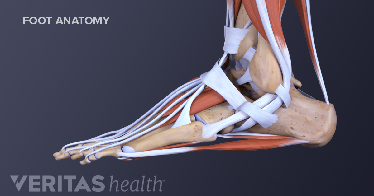
The pain may be substantial resulting in the alteration of daily activities. Plantar fasciitis anatomy the human foot looks simple enough from the outside but its actually pretty complicated.
It contains 19 muscles 26 bones 37 joints 107 ligaments and numerous tendons.
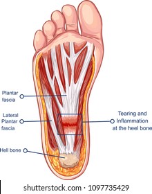
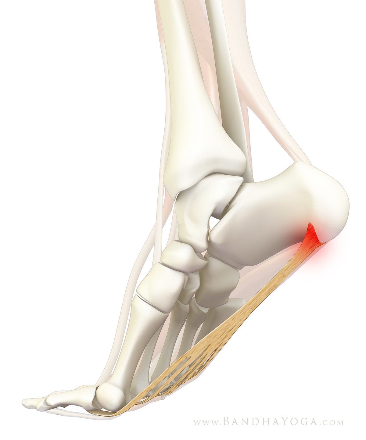
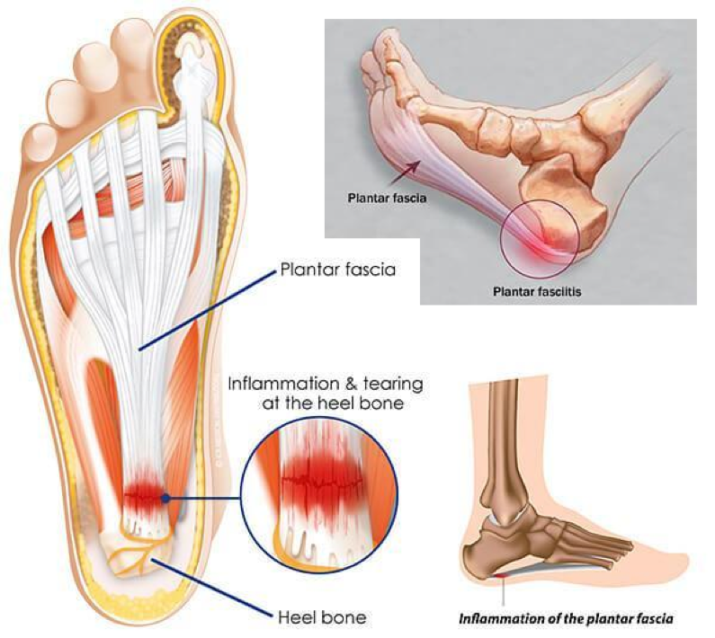
Plantar fascia anatomy. The plantar aponeurosis also known as the plantar fascia is a strong layer of white fibrous tissue located beneath the skin on the sole of the foot. Plantar fasciitis is the pain caused by degenerative irritation at the insertion of the plantar fascia on the medial process of the calcaneal tuberosity. The plantar fascia or plantar aponeurosis forms part of the deep fascia of the sole of the foot and provides a strong mechanical linkage between the calcaneus and the toes.
It runs from the tuberosity of the calcaneus heel bone forward to the heads of the metatarsal bones the bone between each toe and the bones of the mid foot. It denotes the layer of fibrous connective tissue that surrounds different organs muscles bones blood vessels and nerves. Anatomy of the plantar fascia.
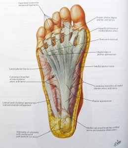
These fibres are mostly longitudinal but also transverse. Anatomy of the plantar fascia the plantar fascia is a complex structure that extends from the medial calcaneal tubercle the heel bone to the proximal phalanges of the toes the bone at the base of the toe at the metatarsophalangeal mtp joints. The word fascia comes from latin meaning a band.
The plantar fascia is the thick connective tissue aponeurosis which supports the arch on the bottom plantar side of the foot. The plantar fascia is the fibrous tissue layer on the plantar surface of the foot that connects the heel bone to the toes. The plantar fascia or plantar aponeurosis is a dense collection of collagen fibres on the sole plantar surface of the foot.
Arising predominantly from the calcaneal tuberosity the plantar fascia attaches distally through several slips. Posteriorly it attaches to the medial process of the tuberosity of the calcaneus proximal to flexor digitorum brevis. Towards the front of the foot at the.
 Extensor Digitorum Longus Muscle An Overview
 Plantar Fasciitis Thermoskin Supports And Braces For
 Botulinum Toxin Treatment Of Plantar Fasciitis Plantar
 Mayo Clinic Q And A Treating Plantar Fasciitis Mayo
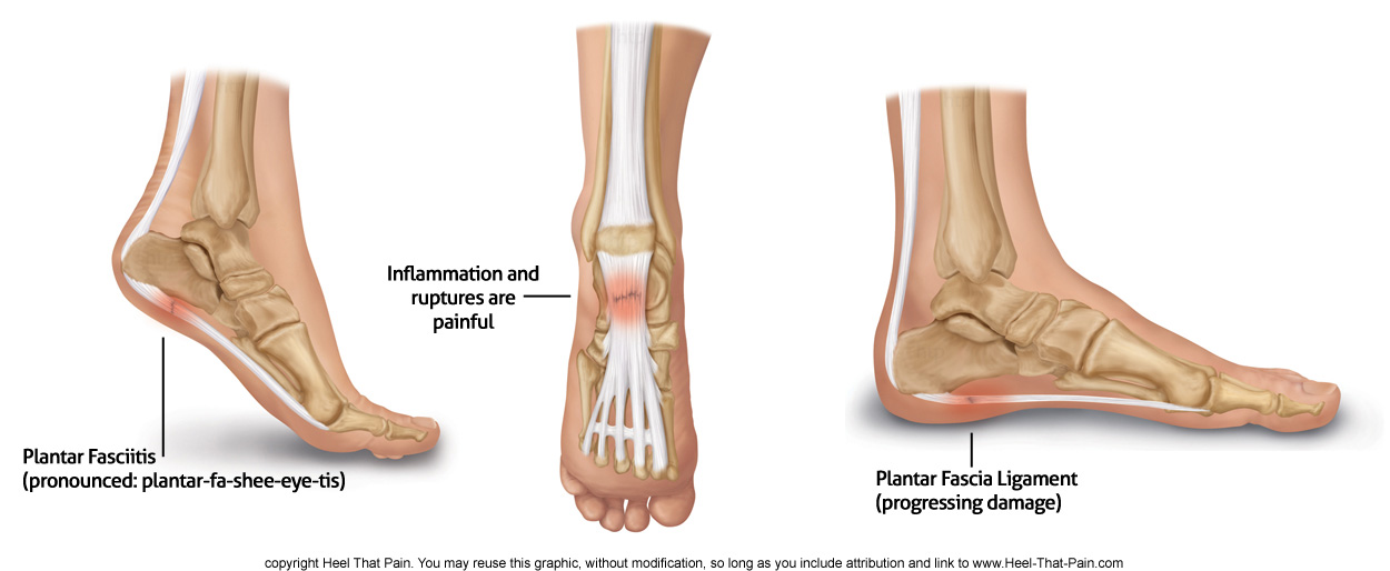 Plantar Fascia Ligament Pain Conditions Heel That Pain
 Pin On Motivation For Power
Plantar Fasciitis Images Stock Photos Vectors Shutterstock
 Running Writings Injury Series Plantar Fasciitis In
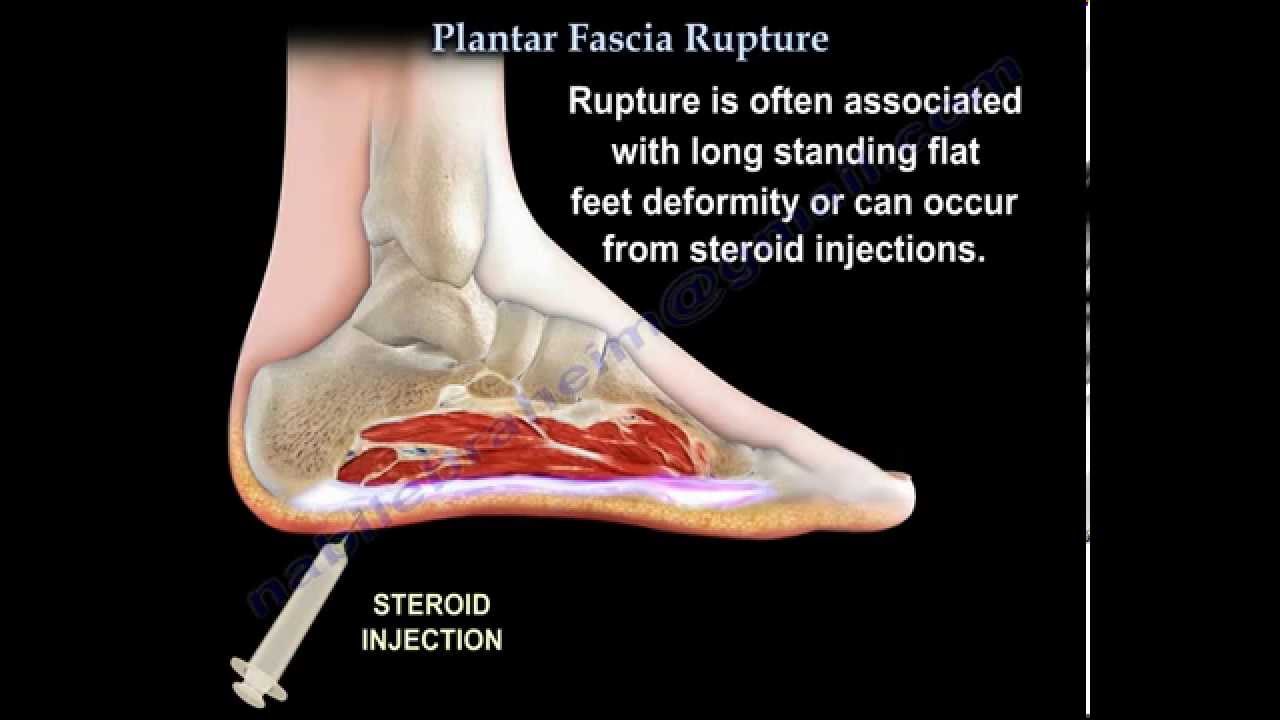 Plantar Fascia Rupture Everything You Need To Know Dr Nabil Ebraheim
 Plantar Fascia Archives Yogi Anatomy
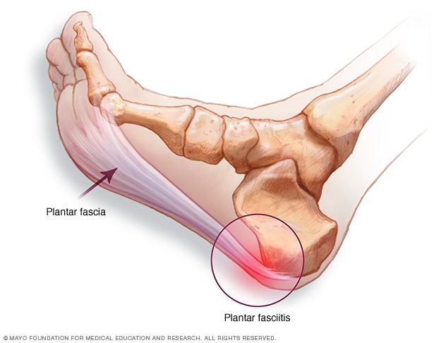 Plantar Fasciitis Symptoms And Causes Mayo Clinic
 Plantar Fasciitis

 Plantar Fasciitis Fleet Feet Columbus
 Plantar Fascia Bottom View Healthlink Bc
 Current And Emerging Concepts In Treating Plantar Fasciitis
Plantar Fasciitis And Bone Spurs Orthoinfo Aaos
 Understanding Plantar Fasciitis Video 1 The Anatomy Of The Plantar Fasciitis
Plantar Fasciitis And Bone Spurs Orthoinfo Aaos
 Plantar Fasciitis Houston Sugar Land Pearland Katy
 Plantar Fasciitis And Heel Spurs Sierra Pacific Orthopedics
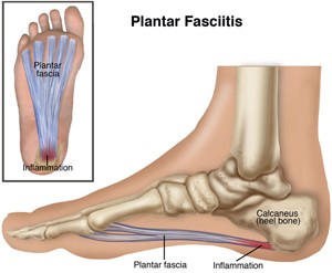
 Plantar Fasciitis Symptoms
 Plantar Fasciitis Is There A Cure Yes Runners Edge

 Plantar Fascia Netter Foot Anatomy Yoga Anatomy Anatomy
 Plantar Fasciitis Overcoming Heel And Arch Pain Naturally
 Plantar Fasciitis Anatomy Body Disease En Fasciitis
 Acland S Video Atlas Of Human Anatomy Plantar Fascia
 Plantar Fasciitis Treatment Relief For Plantar Fasciitis
 My Foot Hurts Plantar Fascia Pain Spinalsymmetry
 The Real Risks Of Steroid Injection For Plantar Fasciitis
 Ouch Why Does My Foot Hurt Synapse
Belum ada Komentar untuk "Plantar Fascia Anatomy"
Posting Komentar