Tracheostomy Anatomy
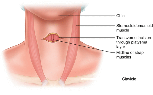
The innermost layer of the trachea consisting of ciliated pseudostratified columnar epithelium and lamina propria a thin layer of connective tissue is covered with a sticky mucus coating produced by the goblet cells present in the region 1. A small hole is cut in the front of the trachea through an incision in the neck.
 Need For Tracheal Humidification Health Products For You
Course of the trachea.

Tracheostomy anatomy. How a tracheostomy is performed surgical anatomy. Contains glands small arteries nerves lymph vessels and elastic fibers trachealis. The trachealis muscle encircles the trachea completely but is most prominent posteriorly due to the lack of cartilage 4.
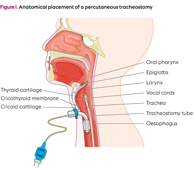
The trachea extends from the inferior margin of the cricoid cartilage. To perfrom a tracheostomy knowledge of the following is required. Anatomy of trachea tracheostomy 1.
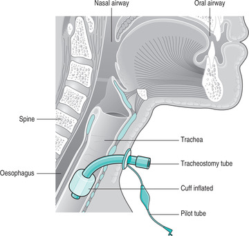
Structure of the tracheal rings. A tracheostomy is a surgical procedure in which an endotrachealtube is inserted directly into the trachea. Components of the larynx.
Nerves vessels and organs. They are the same as a routine open operative tracheostomy with particular. A regimented approach to preparation and performance.
Trachea anatomy and structure tracheal tissues and membranes respiratory mucosa. Trachea anatomy what is the function of the trachea the larynx is a cartilaginous chamber about 4 cm 15 in long figure 1. The trachea is a tube shaped structure consisting of 15 to 20 d shaped cartilage rings anterolaterally bridged by annular ligaments.
Average cross sectional area of the male adult trachea is approximately 28 cm2 transverse. U shaped trachea 27. It is used when intubation through the nose or mouth with an endotracheal tube is not possible or when long term ventilator support is needed such as when a person experiences prolonged unconsciousness and coma.
Its primary function is to keep food and drink out of the airway but it evolved the additional role of sound production phonation in many animals. The superior thyroid notch cricoid and suprasternal notch usually can be easily. Hence we colloquially think of it as the voice box.
Airway Medical Malpractice Part Iii Of Iii Failure To

 Anatomy For Tracheostomy Litfl Medical Blog Ccc Airway
 Educational Products Passy Muir
 Tracheostomy Care Adapted From Various Resources See
 Tracheostomy Practical Slp Info C
 Free Art Print Of Tracheostomy Tube Placement
 Tracheostomy Technique Approach Considerations
 Care Of The Critically Ill Patient With A Tracheostomy
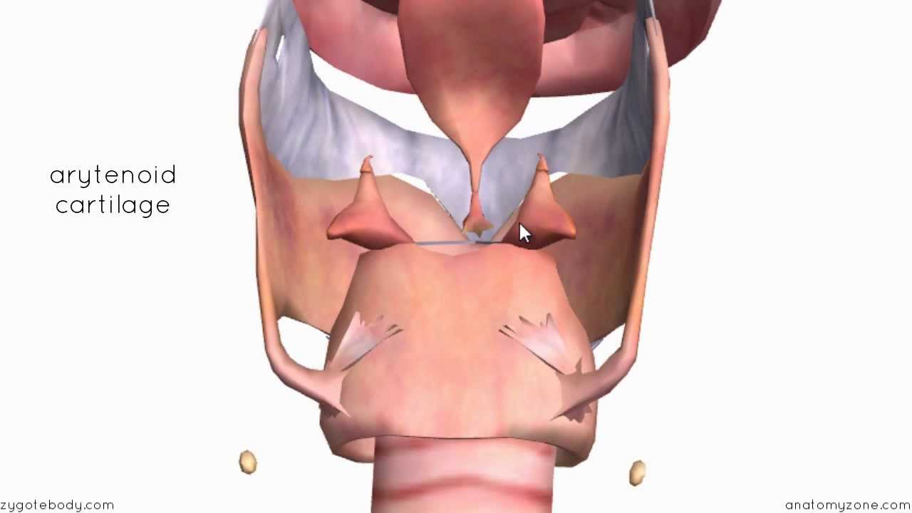
 Anatomy Of Trachea Tracheostomy
 Tracheostomy Tubes Using A Speaking Valve Fact Sheet
 Position Of Tracheostomy Cannulas In The Trachea A An
 Tracheotomy And Tracheostomy Tube How They Help Your Child
 How A Tracheostomy Is Performed
 15061 01x Normal Tracheostomy Tube Placement Anatomy Exhibits
 Anatomy Of Trachea Tracheostomy
 Crash Course In Tracheostomies
 42 Tracheostomy Care Nurse Key
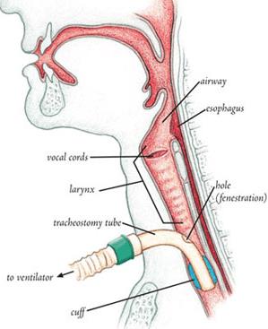
 Tracheostomy With And Without A Ventilator Newport Beach
 Tracheostomy And Cricothyroidotomy Anesthesia Key
 Pin On Respiratory
Tracheostomy
 Anatomy Of Trachea Tracheostomy
 Tracheostomy Pediatrics Clerkship The University Of Chicago
 Anatomy Of Trachea Tracheostomy
 Tracheostomy Basics
 Tracheostomy Stock Photos Tracheostomy Stock Images Alamy

 Tracheostomy Tube Or Stoma Your New Airway
 Figure 2 From Resuscitating The Tracheostomy Patient In The
Belum ada Komentar untuk "Tracheostomy Anatomy"
Posting Komentar