Heart Anatomy External
You can identify the front of the heart by locating the interventricular sulcus and the large pulmonary artery. A trivia quiz called external heart anatomy.
 Functional Anatomy Of The Cardiovascular System Clinical Gate
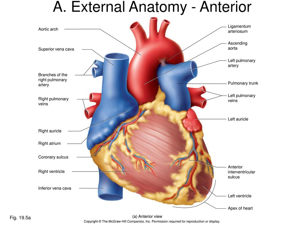
Apex the distal end of the heart that points downward and to the right coronary artery first arteries to branch off of the aorta.

Heart anatomy external. The heart sits within a fluid filled cavity called the pericardial cavity. The walls and lining of the pericardial cavity are a special membrane known as the pericardium. Apex the distal end of the heart that points downward and to the right coronary artery first.
The remainder is supplied by the coronary vasculature which is primarily embedded in the pericardial fat on the surface of the heart and supplies predominantly the epicardium. Heart anatomy external the endocardium and subendocardial tissue receive oxygen and nutrients by diffusion or microvasculature directly from the chambers of the heart. It uses rhythmic electrical impulses that cause the ventricles to contract and force the blood out of the heart so that the returning blood maybe return in a circular fashion.
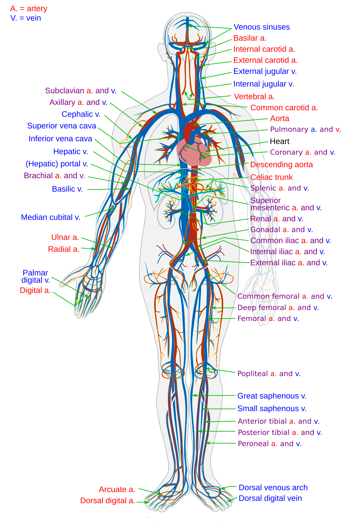
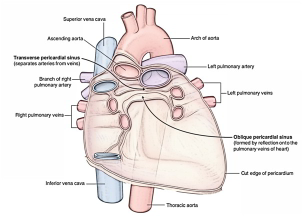
The heart an image of the heart with blank labels attached the circulatory system upper body image with blank labels attached the circulatory system lower body image with blank labels attached the circulatory system a pdf file of the upper and lower body for printing out to use off line. Anatomy of the heart pericardium. Superior vena cava.
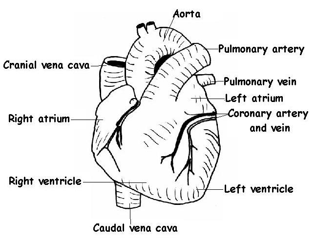
There is a superficial leaf like extension of the atria near the superior surface of the heart one on each side called an auriclea name that means ear likebecause its shape resembles the external ear of a human figure 5. The right one. Heart external anatomy view of the vental surface of the heart.
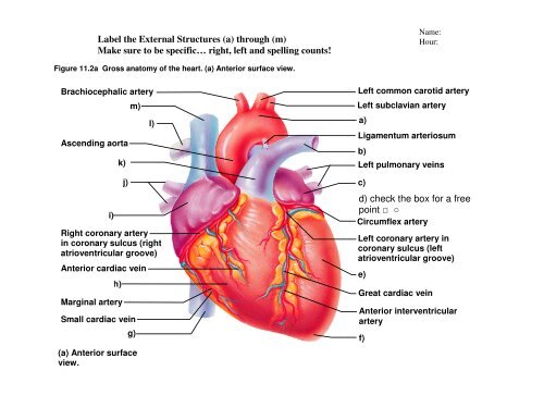
The heart is a muscular organ about the size of a fist located just behind and slightly left of the breastbone. In this article the major anatomy of the heart will be discussed such as the borders surfaces the chambers and the great vessels. Test your knowledge about external heart anatomy with this online quiz.
Because the heart points to the left about 23 of the hearts mass is found on the left side of the body and the other 13 is on the right. Anatomy of the heart external and internal structures duration. Right coronary artery.
Auricles are relatively thin walled structures that can fill with blood and empty into the atria or upper chambers of the heart.
 Anatomy Of The Heart Review
 The Anatomy And Physiology Of Animals Heart Worksheet
 Chapter 19 The Circulatory System The Heart Ppt Download
 Blood Vessel Wikipedia
 2 External Features Of The Heart
 External Imaging Aneterior Atlas Of Human Cardiac Anatomy
 Easy Notes On Heart Learn In Just 4 Minutes Earth S Lab
 External Features Of Heart
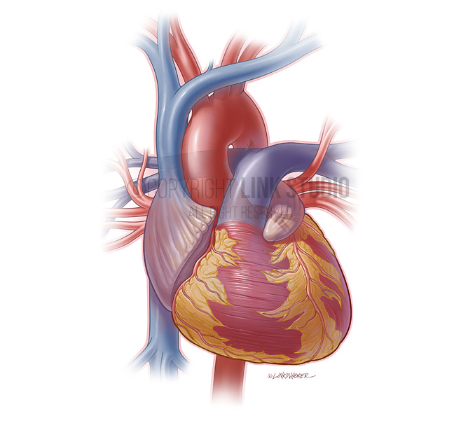 Heart Anatomy Link Studio
 External Heart Anatomy Posterior View Diagram Quizlet
Lab 04 Heart Anatomy
 Anatomical Diagrams Of Heart Heart Failure Online
 Coronary Artery Anatomy
 Human Anatomy Heart Power Point
 2 External Features Of The Heart
 Heart External Anatomy Illustration Stock Image C027
 Heart Gross Anatomy Practice Quiz
 2 External Features Of The Heart
Circulatory Systems In Animals Transport Systems In
 Diagram External Structure Human Heart Seen Stock Vector
 The External Anterior Anatomy Of The Heart Showing Coronary Arteries
 Heart Wikipedia
 The Heart
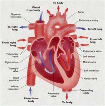
The Cardiovascular System The Heart

Belum ada Komentar untuk "Heart Anatomy External"
Posting Komentar