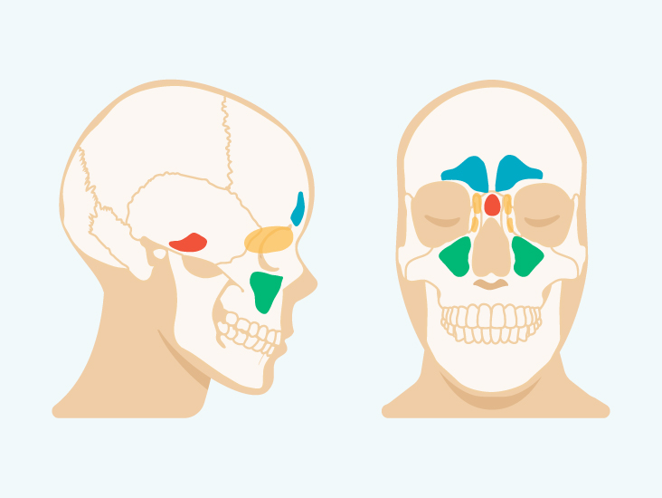
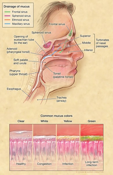
Anatomy Of Sinus Drainage
A thin wall called the septum divides the nose. Anterior middle and posterior.
 Sinus Cavities In The Head Anatomy Diagram Pictures
The inferior sagittal and straight sinuses are exemptions to this rule.

Anatomy of sinus drainage. Most individuals have four paired cavities located in the cranial bone or skull. Armando hasudungan 297486 views. There are three ethmoidal sinuses.
The relationships of this sinus are of clinical importance the pituitary gland can be surgically accessed via passing through the nasal roof into the sphenoid sinus and through the sphenoid bone. Figure 1 provides an overview of the drainage pathways of the paranasal sinuses that are based upon the skull illustrated in figures 3 and 4. The frontal ostium extends between the anterior and posterior walls of the frontal sinus is demarcated by a variably shaped ridge of bone on the anterior wall of.
Anatomy of the nasal cavitymov. Generally the walls of these drainage pathways are formed by visceral periosteum and dural reflection both lined with endothelium. Normally these structures help humidify and filter air.
Their drainage path may vary depending on how the frontal sinus drainage pathway fsdp develops 12. They empty into the nasal cavity at different places. As long as your sinuses are draining properly the increase in mucus isnt a problem although it can be annoying.
Anatomy of the nasal cavitymov. The inside of the nose has ridges called turbinates. Each frontal sinus narrows down to an inferior margin designated the frontal ostium figs 1 and 3d f.
The frontal sinus has the most complex and variable drainage of any paranasal sinus. Instead they drain to the dural sinuses which subsequently drain to the internal jugular vein. Sinus drainage can be worsened by a number of factors such as.
Skip navigation sign in. Most of the sinuses drain into the nose through a small channel or drainage pathway that doctors call the middle meatus. The sphenoid sinuses drain out onto the roof of the nasal cavity.
Sinus anatomy in common usage sinus usually refers to the paranasal sinuses which are air cavities in the cranial bones especially those near the nose and connecting to it. Clinical anatomy nasal cavity and sinuses duration. The real problems start when the passages become blocked or fail to let the sinuses drain effectively.
The two frontal sinuses drain through the frontonasal duct which opens in the lateral wall of the nasal cavity at the semilunar hiatus 2. The resulting pressure can be intolerable for many people.
 Cerebral And Sinus Vein Thrombosis Circulation
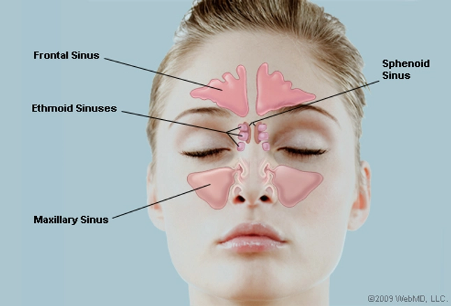 What Are The Sinuses Pictures Of Nasal Cavities
 Anatomy Notes Balloon Sinuplasty
 Paranasal Sinuses Wikipedia
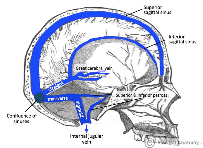 Venous Drainage Of The Cns Cerebrum Teachmeanatomy
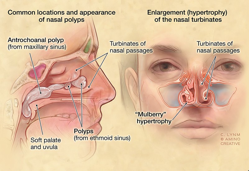
 Nasal Cavity
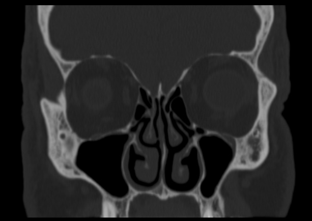
Startradiology
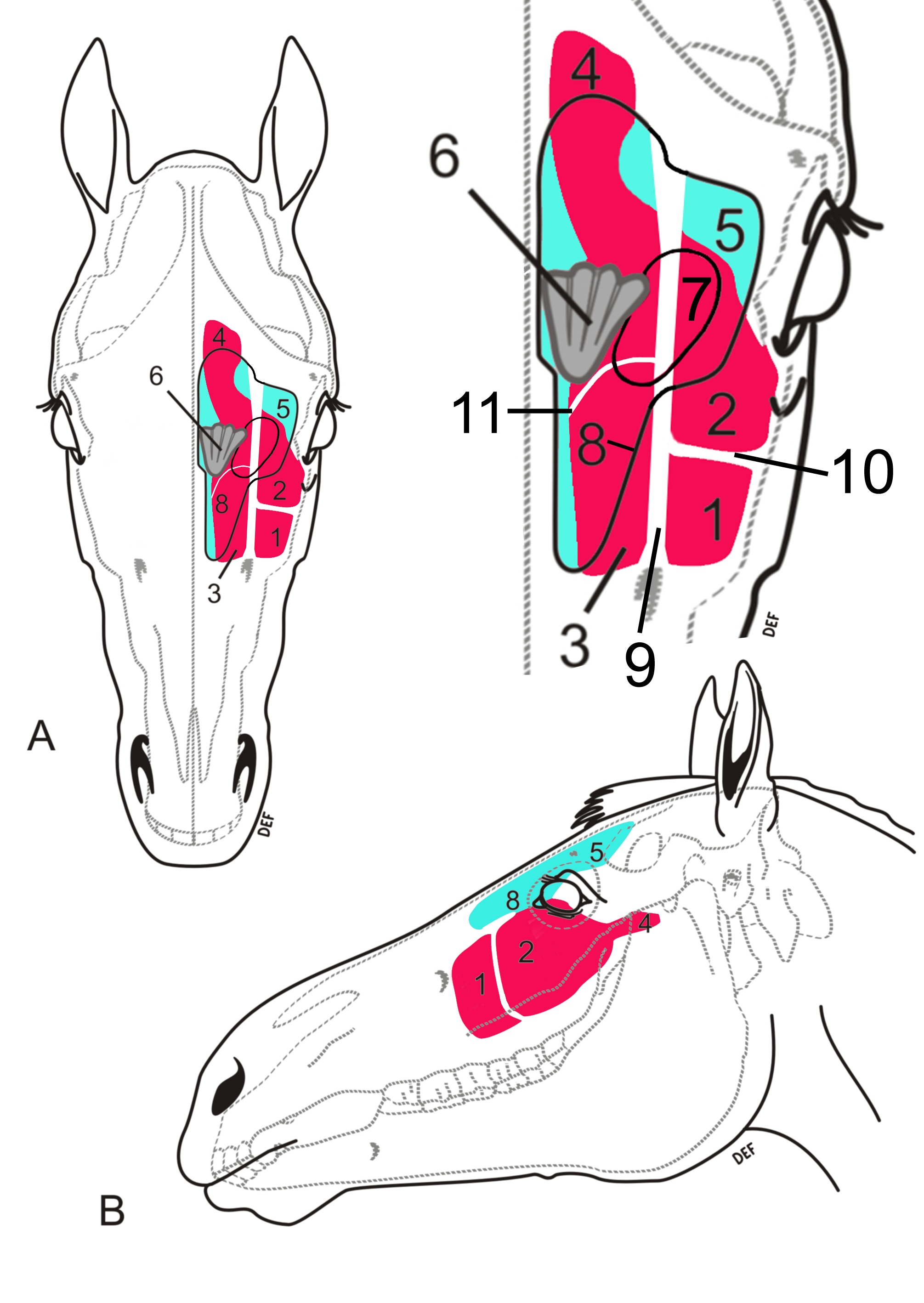 Equine Sinus Conditions Large Animal Hospital College Of


 Balloon Sinus Dilation
 Equine Sinus Disease A Hidden Danger Expert How To For
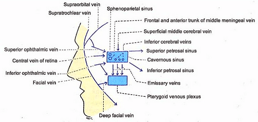
 Simplified Anatomy Of The Eye Paranasal Sinuses And Venous
 Sinuses Picture Image On Medicinenet Com
 Applied Anatomy Of Cavernous Sinus Epomedicine
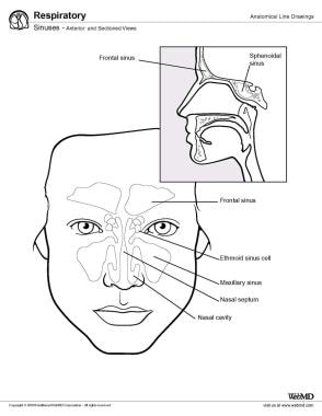 Paranasal Sinus Anatomy Overview Gross Anatomy
 Dr Reuben Setliff Pioneer In Sinus Care
 Startradiology
 Chronic Sinusitis Allergy Asthma And Sinus Specialists

 Nasal Sinus Program Davis Ear Nose Throat Sleep Center
 Dr Reuben Setliff Pioneer In Sinus Care
 Clinical Anatomy Nasal Cavity And Sinuses Youtube
Belum ada Komentar untuk "Anatomy Of Sinus Drainage"
Posting Komentar