Calcaneus Anatomy
From the latin calcaneus or calcaneum meaning heel or heel bone is a bone of the tarsus of the foot which constitutes the heel. The rear half of the heel bone is known as the tuber calcanei.

The calcaneus provides insertion points for the abductor hallucis and.
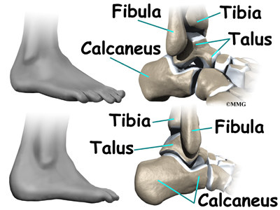
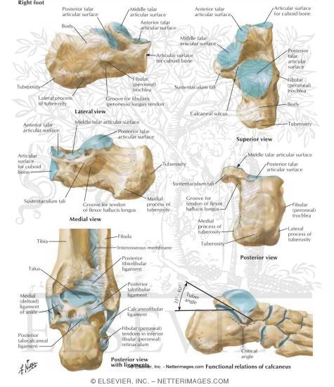
Calcaneus anatomy. The calcaneus is an irregular roughly box shaped bone sitting below the talus and its anterior aspect is inclined cranially. It is responsible for the visible projection of the foot that constitutes the heel. Talussmall foot bone that works as a hinge between the tibia and the fibula together the calcaneus and the talus form the subtalar joint.
Structure of calcaneus anterior surface. The calcaneus has a unique design and structure. At the front the heel bone features many curves to accommodate the talus and the many different tarsal bones which lead to the metatarsals and phalanges that make up the front of the foot and toes.
The calcaneus connects with the talus and cuboid bones. The inferior or plantar surface is wider posteriorly and convex from side to side. The subtalar joint allows side to side movement of the hindfoot and is especially important for balance on uneven surfaces.
In humans the calcaneus kælˈkeɪniəs. The heel bone is the largest bone in the foot. Case discussion calcaneal fractures and other pathology are common and thus it is important to have a detailed understanding of calcaneal anatomy.
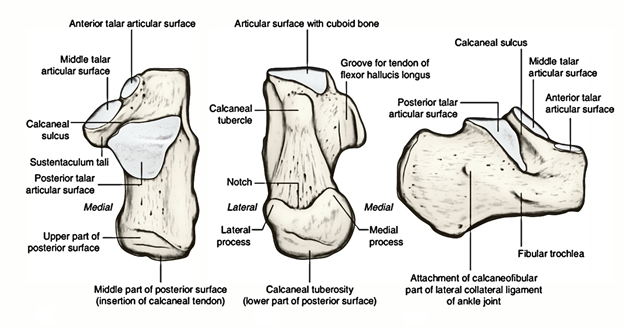
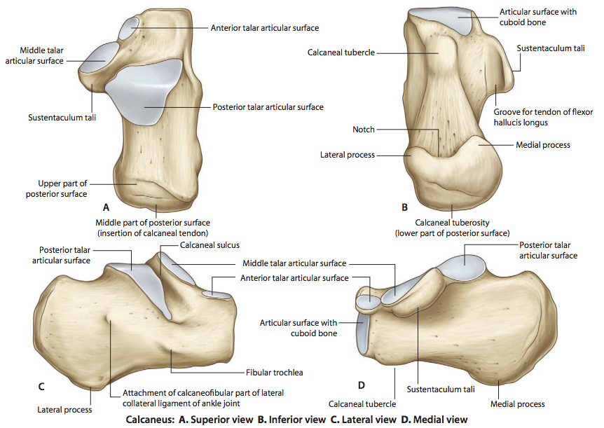
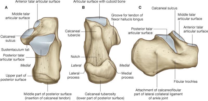
Of all of the bones in the foot the heel bone is the largest. The calcaneus is an irregular bone cuboid in shape whose superior surface can be. The superior calcaneal surface of the calcaneus has 2 parts.
The connection between the talus and calcaneus forms the subtalar joint. In some other animals it is the point of the hock. Muscle and ligament attachments.
Two muscles of the foot abductor hallucis and abductor digit minimi extend from the heel bones sides. As the calcaneus is the largest of the bones in the foot. The calcaneus also called the heel bone is a large bone that forms the foundation of the rear part of the foot.
The anterior surface is the smallest surface of the bone. The anatomy of the calcaneus is outlined as indicated above.
 Figure Calcaneus Anatomy Contributed By David R Davis Md
 Calcaneus Anatomy
 Calcaneus Heel Bone Fractures Orthoinfo Aaos
 Anatomy Of The Calcaneus Calcaneus
 Easy Notes On Calcaneus Learn In Just 4 Minutes
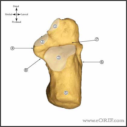
 Calcaneus Anatomy Eorif
 Anatomy Of The Calcaneus Everything You Need To Know Dr Nabil Ebraheim
 Calcaneus Approach Sinus Tarsi Approach Ao Surgery
 Calcaneus
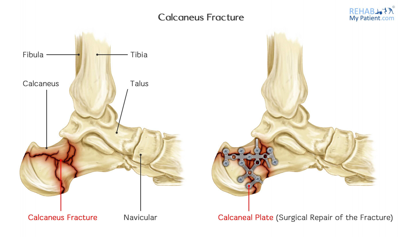 Calcaneus Fracture Rehab My Patient
Calcaneus Heel Bone Fractures Orthoinfo Aaos
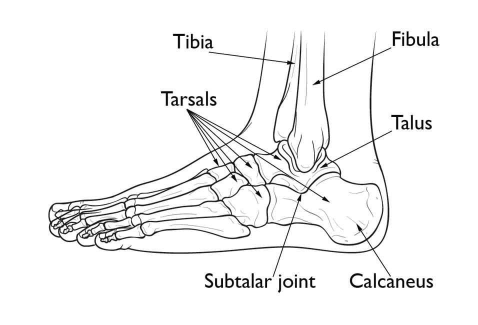
 Ankle Foot Anatomy
Tarsal Coalition Orthoinfo Aaos
Ankle Anatomy Orthogate
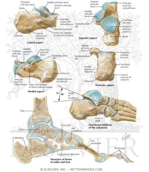
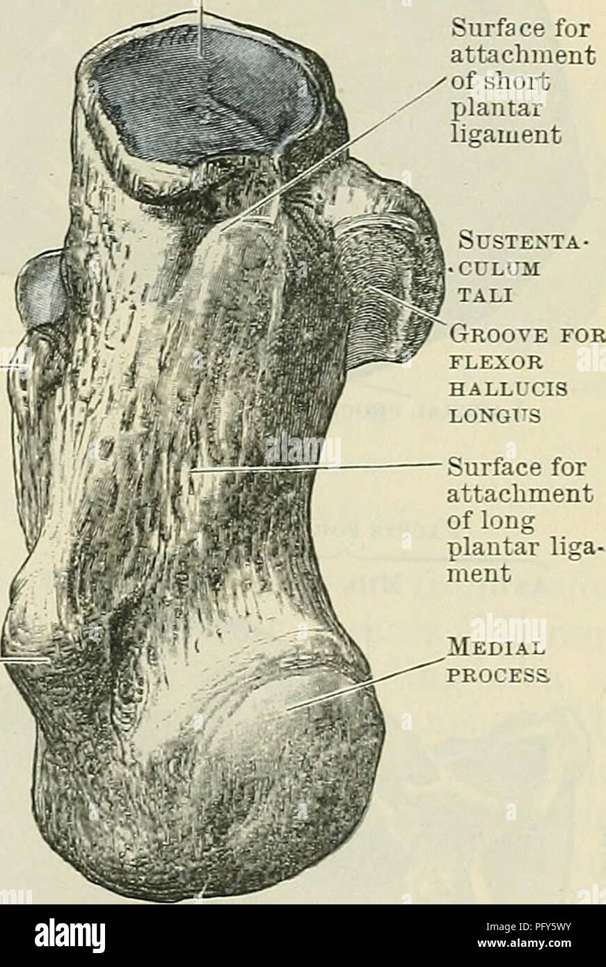
 Calcaneus Anatomy And Attachments Bone And Spine
 Cunningham S Text Book Of Anatomy Anatomy Tuberosity A Fig
 Anatomy Archives Page 5 Of 10 Bone And Spine
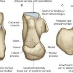
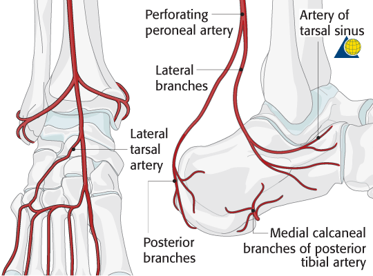
 The Subtalar Joint Ligaments Neurovascular Teachmeanatomy
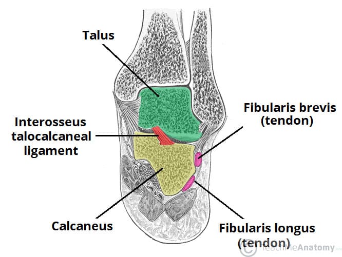
![]() Calcaneus Anatomy And Pathology Kenhub
Calcaneus Anatomy And Pathology Kenhub
 Intra Articular Tongue Type Fractures Of The Calcaneus
 Get To Know The Ankle Joint Yoga Journal
 Foot And Ankle Musculoskeletal Key
 Calcaneus Radiology Reference Article Radiopaedia Org
 Achilles Tendon Anatomy And Importance
 Calcaneus Anatomy Eorif
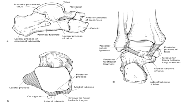
 Talus Bone Wikipedia
 Calcaneus Anatomy And Attachments Bone And Spine
 Core Knowledge Calcaneal Fractures St3 Orthopaedic
![]() Calcaneus Anatomy And Pathology Kenhub
Calcaneus Anatomy And Pathology Kenhub
Belum ada Komentar untuk "Calcaneus Anatomy"
Posting Komentar