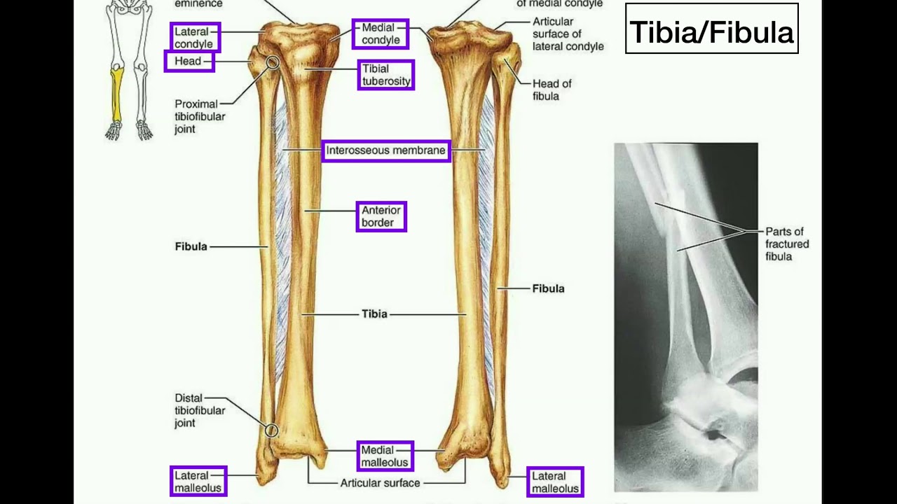
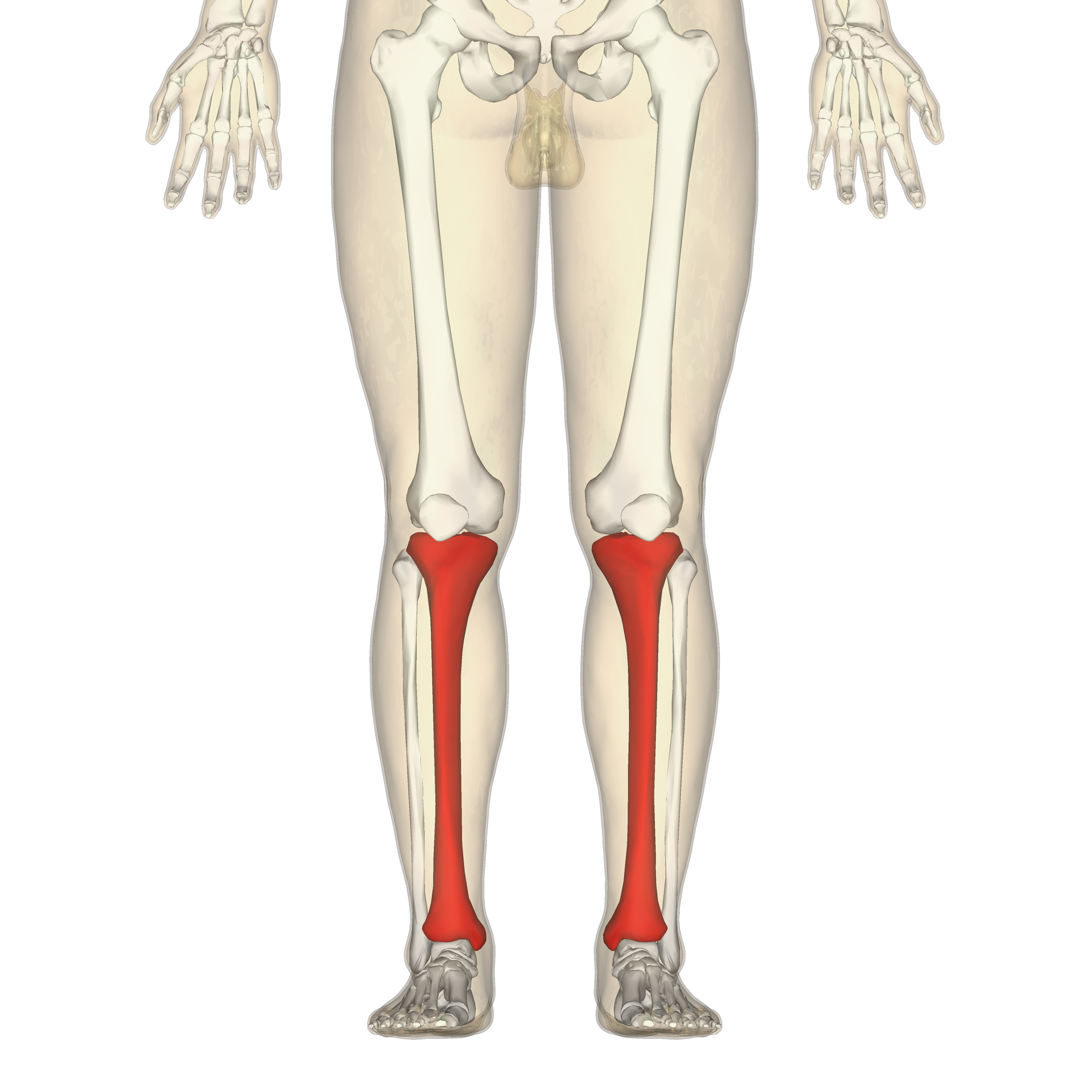
Tibial Plafond Anatomy
Soft tissues very poor thin skin absence of muscle and adipose tissue lack of deep veins. Males 3 x.
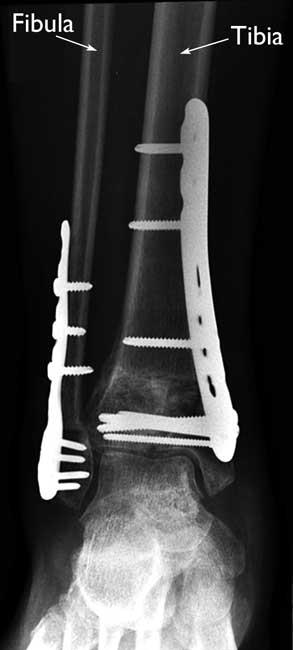
 Tibial Pilon Fracture With Intact Fibula Everything You Need To Know Dr Nabil Ebraheim
Up to 50 incidence of associated injuries.
Tibial plafond anatomy. Tibial plafond fracture orif with anterolateral approach and plate fixation ankle and hindfoot ankle simple bimalleolar fracture orif with 13 tubular plate and cannulated screw of medial malleol. Plafond fractures are infrequent injuries accounting for 7 10. Tibial plafond fractures introduction.
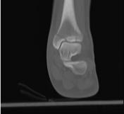
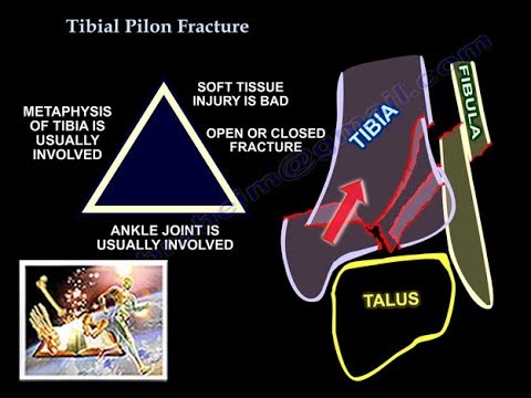
Fracture of tibial weight bearing surface due to axial compression. It involves the articular surface of the ankle joint. 1 a pilon fracture also called a plafond fracture is a fracture of the distal part of the tibia involving its articular surface at the ankle joint.
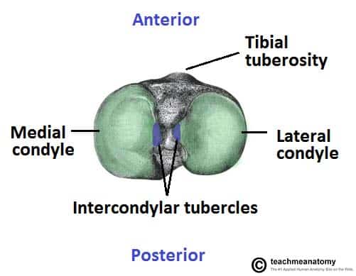
11 originally proposed an anatomical nine zone grid scheme to the articular surface of the talus to more easily describe the location of an ocl and have also applied this to the articular surface of the distal tibial plafond figure 1. First branch of popliteal artery. Mechanism typically occurs as a result of an axial loading injury which drives the talus into the tibial.
Rapid axial load very high energy. Passes between 2 heads of tibialis posterior and interosseous membrane iom. The distal portion of the tibia is known as the plafond which.
Clinical features of pain swelling deformity and crepitus about the. Especially vunerable over anteromedial tibia. 35 40 years.
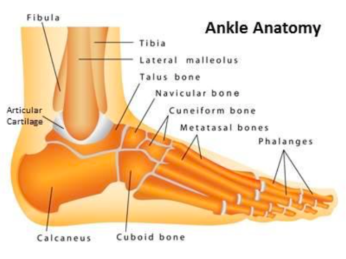
The tibial plafond lateral malleolus and medial malleolus form a mortise a socket in which the talus sits figure 2. These are considered to represent 1 10 of all lower limb fractures 6. It is also known as pilon fracture and explosion fracture.
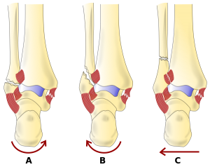
The cause of tibial plafond fracture is axial or rotational forces occurring from motor vehicle accidents or falling from a height. Pilon fractures are caused by rotational or axial forces mostly as a result of falls from a height or motor vehicle accidents. Articulates with the talus and fibula laterally via the fibula notch.
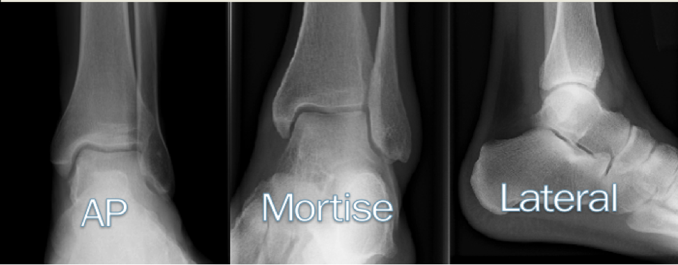
A pilon fracture is a type of fracture involving the distal tibia. Tibial plafond fracture is an uncommon fracture occurring in the distal region of the tibia. Radiographic examination of the ankle bones.
Distal tibia forms an inferior quadrilateral surface and pyramid shaped medial malleolus. Although the ligaments are needed to give the ankle its full stability the bony congruity of the mortise and the talus is a necessary component as well forming the most congruent joint in the lower extremity. Anterior tibial artery.
 The Tibia Proximal Shaft Distal Teachmeanatomy
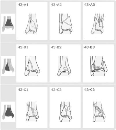
 39 Tibial Plafond Pilon Fractures
 These Four Tibial Plafond Fracture Radiographs Illustrate
 Anatomy 101 Ankle Syndesmosis Distal Tibiofibular Joint
Right Tibia Anatomy Tips Electrical Wiring
 Tibia Wikipedia

 Pilon Fractures Of Tibia Presentation And Treatment Bone
 Ppt Pilon Fractures Powerpoint Presentation Free Download
 Figure 2 From Anatomy Of Pilon Fractures Of The Distal Tibia
 Tibial Plafond Fractures Trauma Orthobullets
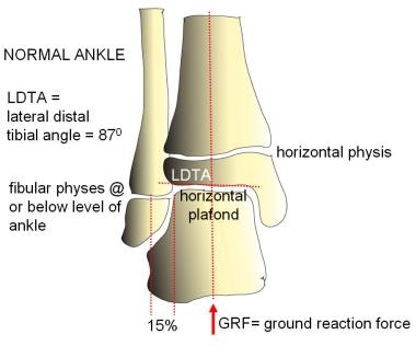 Pediatric Ankle Valgus Background Anatomy Pathophysiology
 Pilon Fracture Radiology Case Radiopaedia Org
 Pilon Fracture Wikipedia
 L11 Pilon
 Pilon Fracture Radiology Reference Article Radiopaedia Org
 Ankle Fractures Core Em
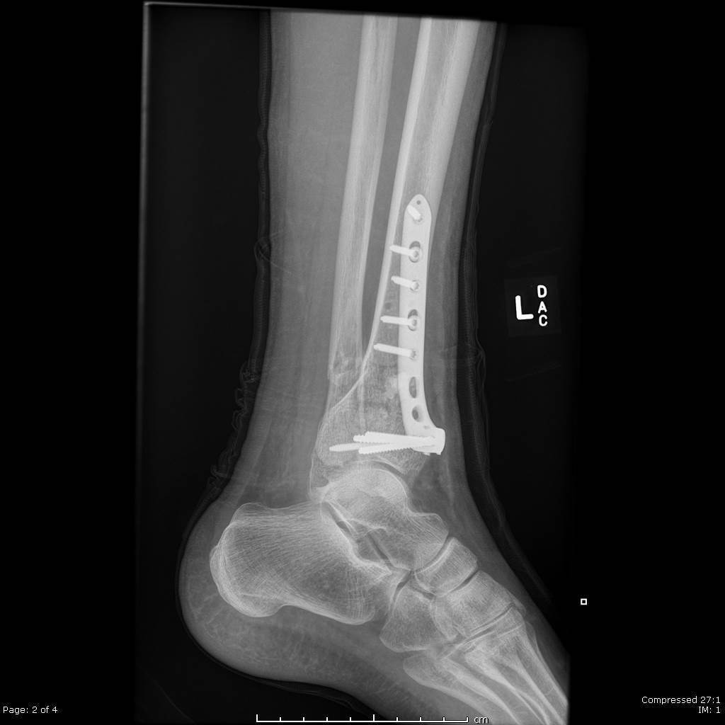
 Treatment Of Malreduced Pilon Fracture A Case Report And
 Tibial Pilon Fracture Everything You Need To Know Dr Nabil Ebraheim
 Pin By Andres Sanchez On Radiology Medical Anatomy
Current Concepts In Trauma Ankle And Pilon Fractures
 Ankle And Foot Fractures Physiopedia
 Pilon Fractures Of The Ankle Orthoinfo Aaos
 Right Tibia Anatomy Tips Electrical Wiring
Belum ada Komentar untuk "Tibial Plafond Anatomy"
Posting Komentar