Female Ureter Anatomy
From the pelvic brim to the bladder. The ureter is 25 30 cm long and has three parts.
Amicus Illustration Of Amicus Anatomy Pelvic Female
As the anatomy of the male and female urogenital wing shows some significant differences the structure of the urethras decisively mirrors them.
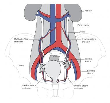
Female ureter anatomy. The female urethra begins at the bottom of the bladder known as the neck. Therefore the male and female urethras are examined separately. One of the most common areas of.
Anatomy of the female urinary system showing the kidneys ureters bladder and urethra. In females the ureters pass through the mesometrium and under the uterine arteries on the way to the urinary bladder. An inset shows the renal tubules and urine.
Next to the ureteric fold the gonadal vessels form an adjacent fold in female infundibulopelvic or suspensory ligament of ovary. Citation needed blood and lymphatic supply. The female urethra is clearly shorter than the male variant with an average length of 3 5 cm.
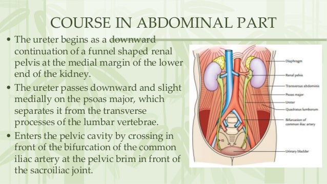
From the renal pelvis to the pelvic brim. It extends downward through the muscular area of the pelvic floor. In anatomy the urethra is a tube that connects the urinary bladder to the urinary meatus for the removal of urine from the body of both females and males.
The uterus is also shown. Before reaching the urethral opening urine passes through the urethral sphincter. In human females and other primates the urethra connects to the urinary meatus above the vagina whereas in marsupials the females urethra empties into the urogenital sinus.
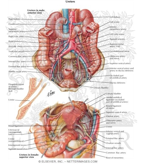
However internally they are similar the kidneys of both men and women for example look and function the same for both genders. As they cross the pelvic brim the ureters are in close proximity to the ovaries. Female urology and external sexual anatomy.
Anatomy and function of the female urethra. But we also differ in some ways too women have much shorter ureters the tube that connects your bladder to your urethra and therefore are at greater risk of bladder infections. They must be identified during pelvic surgery to ensure that they are not accidentally damaged.
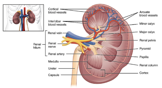
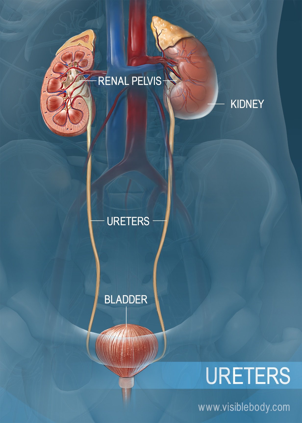
Shows the right and left kidneys the ureters the bladder filled with urine and the urethra. The inside of the left kidney shows the renal pelvis. The anatomical course of the ureters is of surgical importance as they travel close to other structures in the pelvis.
Ureter urinary bladder and malefemale urethrae structures and walls urinary system anatomy duration. Left testicular or ovarian vessels. The ureter has an arterial supply which varies along its course.
Within the bladder wall. Females use their urethra only for urinating but males use their urethra for both urination and ejaculation. Intravesical or intramural ureter.
The upper part of the ureter closest to the kidney is supplied by the renal arteries. The external urethral sphincter is a striated muscle that. Just above the entry to the pelvis the ureter is still covered by peritoneum by virtue of the ureteric fold.
Anatomy of the female urinary system.
 New Paradigms In Endometriosis Surgery Of The Distal Ureter
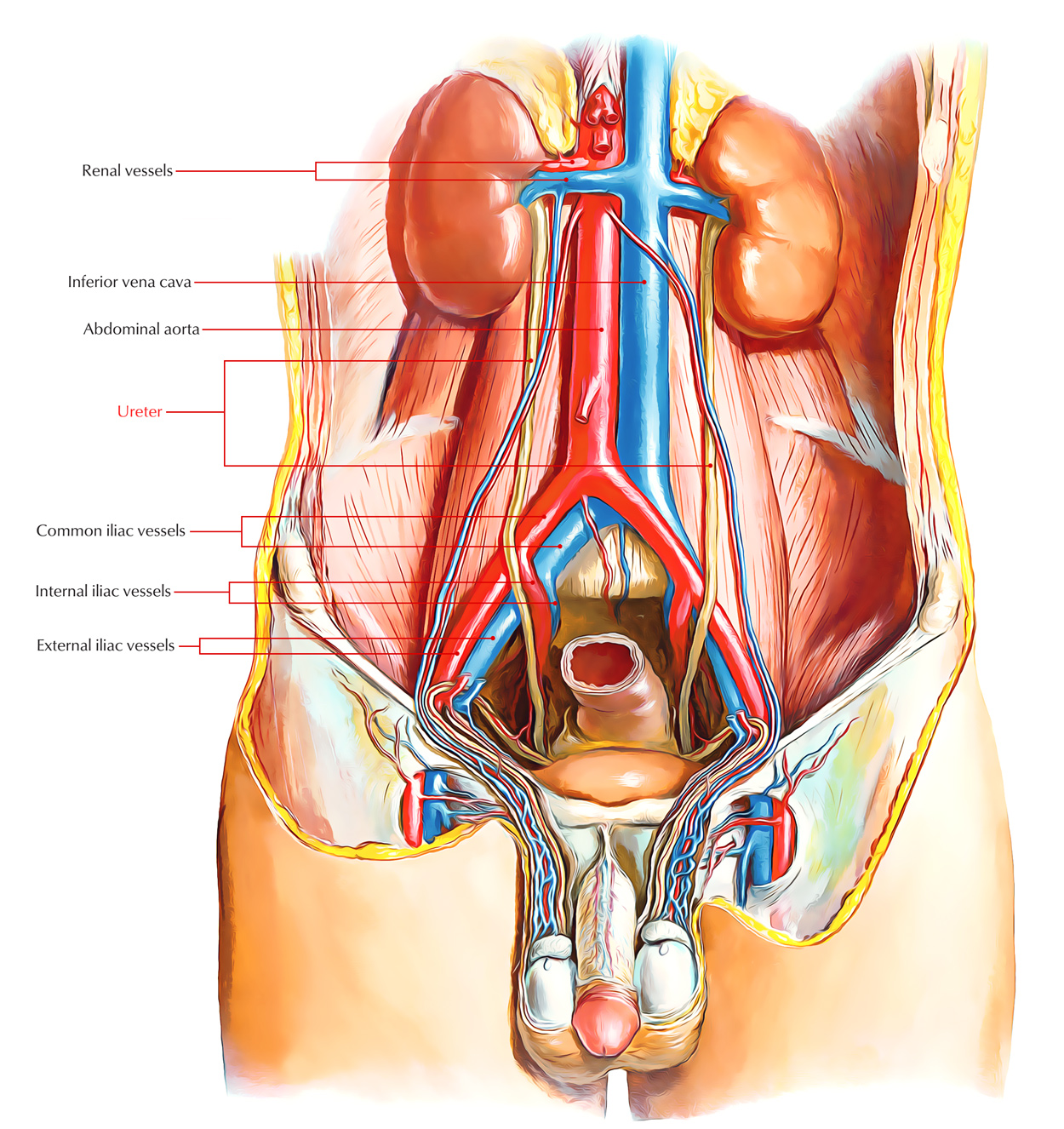 Easy Notes On Ureter Learn In Just 4 Minutes Earth S Lab
:background_color(FFFFFF):format(jpeg)/images/library/11902/renal-cortex_english.jpg) Kidneys Ureters Suprarenal Glands Anatomy Location Kenhub
Kidneys Ureters Suprarenal Glands Anatomy Location Kenhub
 Definition Of Ureter Nci Dictionary Of Cancer Terms

:watermark(/images/watermark_5000_10percent.png,0,0,0):watermark(/images/logo_url.png,-10,-10,0):format(jpeg)/images/atlas_overview_image/385/ZZNxxFMmiwqAMjF09TOQ_ureters-in-situ_english.jpg) Ureters Anatomy Innervation Blood Supply Histology Kenhub
Ureters Anatomy Innervation Blood Supply Histology Kenhub
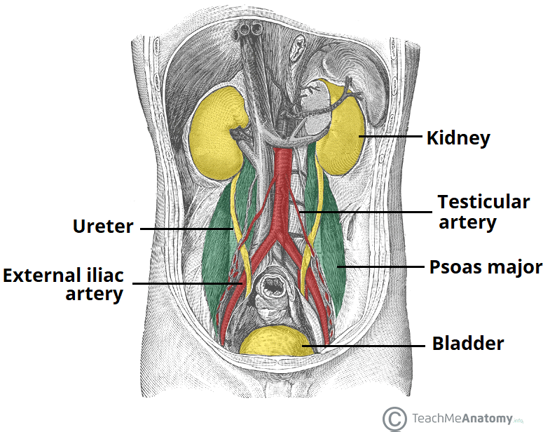 The Ureters Anatomical Course Neurovascular Supply
 Intraperitoneal And Retroperitoneal Anatomy The 3rd
 Renal System Definition Function Diagram Facts
 Anatomy And Normal Microbiota Of The Urogenital Tract
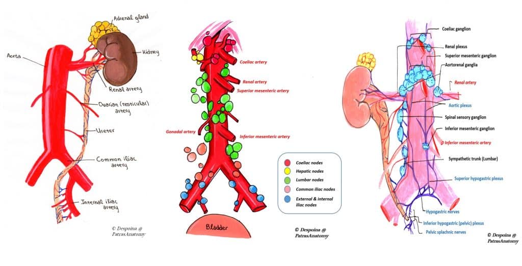 The Ureters Anatomical Course Neurovascular Supply
 Anatomy Of The Female Urinary Tract
 Anatomy Of The Kidney And Ureter Sciencedirect
 Urinary Tract Infections In Women
 Ureter Anatomy Overview Gross Anatomy Microscopic Anatomy
 19 5 Ureters Urinary Bladder And Urethra Biology Libretexts
 Anatomic Relations Of Ureters Ureters

 Bladder Urethra Anatomy Renal Medbullets Step 1


Belum ada Komentar untuk "Female Ureter Anatomy"
Posting Komentar