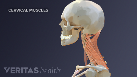
Anatomy Of Cervical Muscles
Cervical spine anatomy there are seven vertebrae in the cervical spine neck area which surround the spinal canal and the spinal cord. Discs made up of gelatinous material act as cushioning between these vertebra with nerves passing out of the spinal canal between the disc and vertebra.
 Nerves Blood Vessels And Lymph Advanced Anatomy 2nd Ed
Therefore for this articles purposes the neck muscles may be divided into four major structural groups each in its own quadrant.

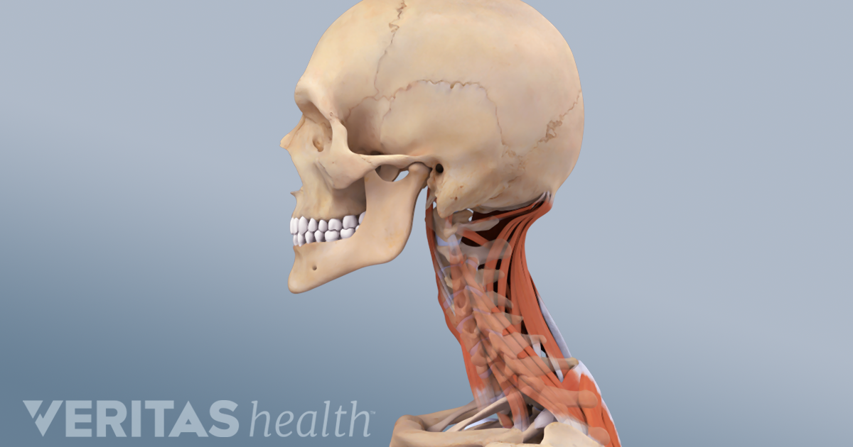
Anatomy of cervical muscles. The musculature of the neck is comprised of a number of different muscle groups. Rectus capitis lateralis originates from the first. The cervical spine straightens or moves directly backward with the chin tilting up.
Together the scalenes act to flex the neck. The muscles in the neck are responsible for the movement of the head in the cervical region in all directions. The posterior scalene also originates from the cervical spine but attaches instead to the second rib.
They move the head in every direction pulling the skull and jaw towards the shoulders spine and scapula. The cervical spine section contains seven vertebrae c 1 through c 7 and eight nerve pairs c 1 through c 8. Head and neck motions typically involve one or more of the following movements of the cervical spine.
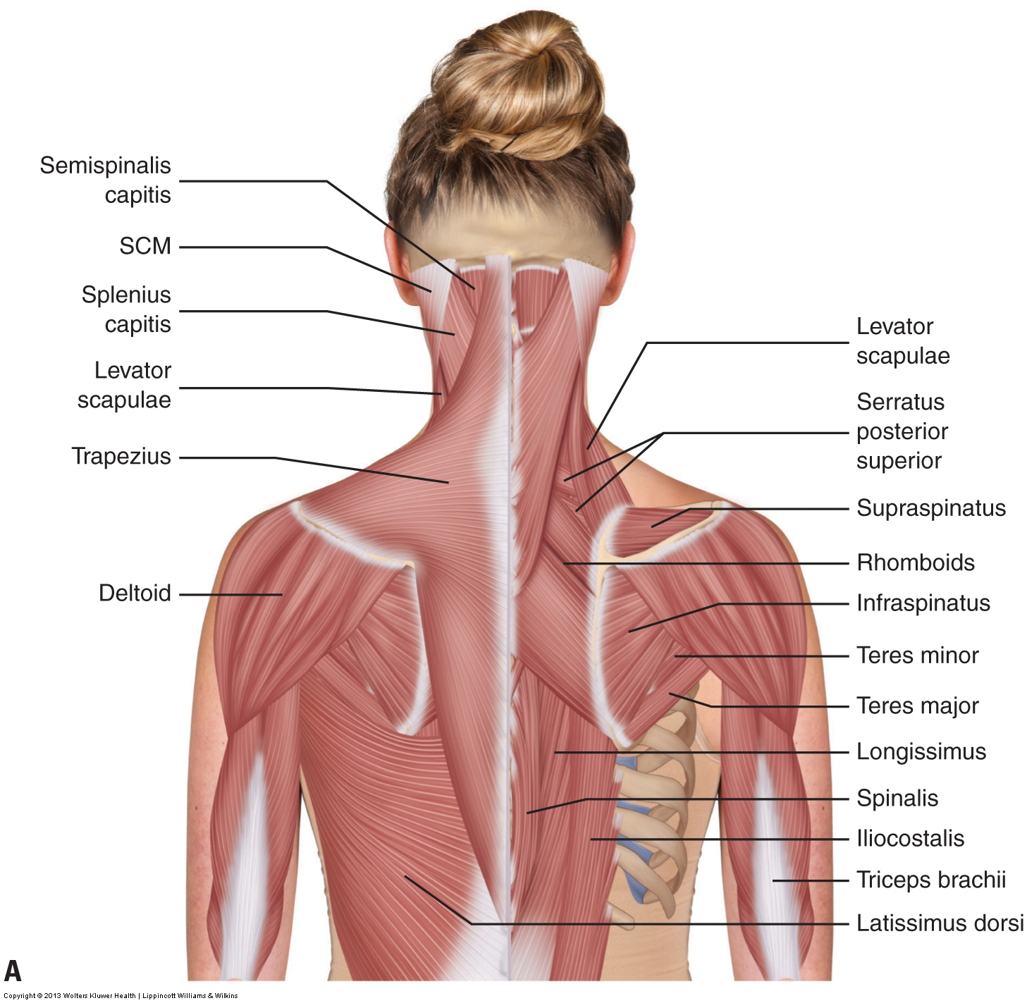
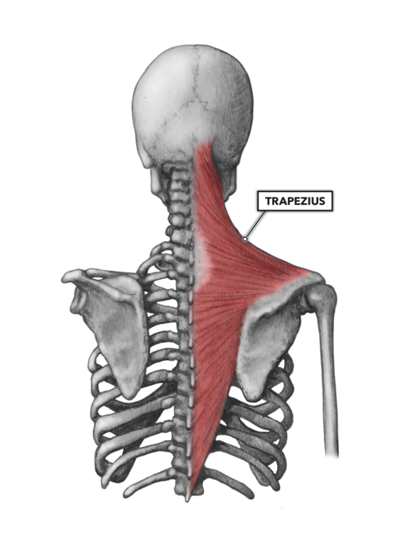
Longus capitis begins between the third and sixth cervical vertebrae. They can also be recruited as accessory muscles of respiration. The fan shaped trapezius muscles extend from the back of the skull down to the middle of the back along the spine and fan over into the shoulders.
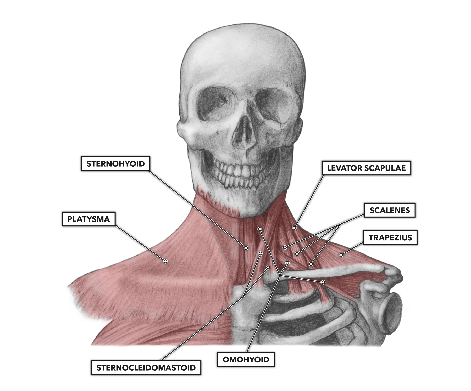
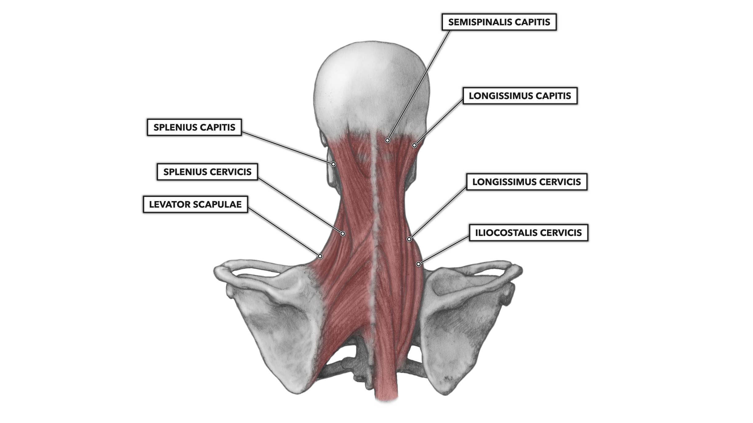
These muscles give the sides of the neck their. These muscles are innervated by the posterior rami of various cervical spinal nerves. The neck muscles including the sternocleidomastoid and the trapezius are responsible for the gross motor movement in the muscular system of the head and neck.
Muscles of the neck. The top section of the spine is the cervical section which contains nerves that innervate muscles of the head neck and thoracic cavity as well as transmit sensory information to the cns. The cervical spine bends directly forward with the chin tilting down.
Rectus capitis anterior begins at the first cervical vertebrae. The musculature of the neck is comprised of a number of different muscle groups. Longus colli begins between the third and sixth cervical vertebrae.
 Crossfit Cervical Muscles Part 1
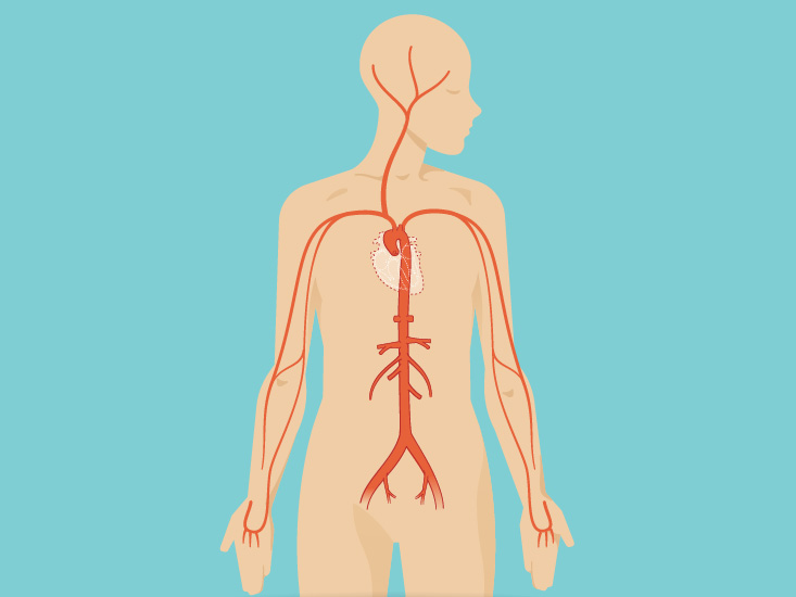 Neck Anatomy Area Diagram Body Maps
 Neck Muscles Anatomy Art Watercolor Splash
 Muscles Of The Neck Musculature Of The Cervical Spine
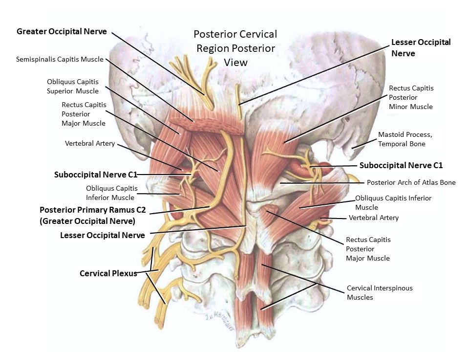
 Suboccipital Nerve An Overview Sciencedirect Topics
A B The Neck Head Musculoskeletal Anatomy Showing The 216
 Cervical Motor Control Part 1 Clinical Anatomy Of Cervical
![]() Neck Anatomy Muscles Glands Organs Kenhub
Neck Anatomy Muscles Glands Organs Kenhub
 Neck Muscles And Other Soft Tissues
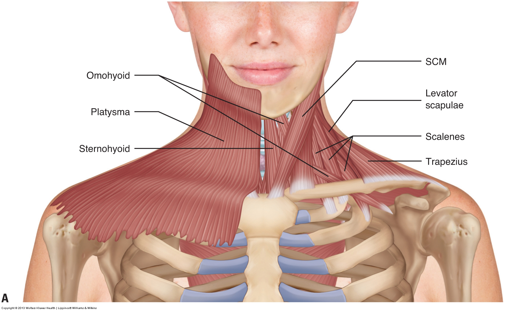
 Anterior Neck Muscles Preview Human Anatomy Kenhub
 Crossfit Cervical Muscles Part 2
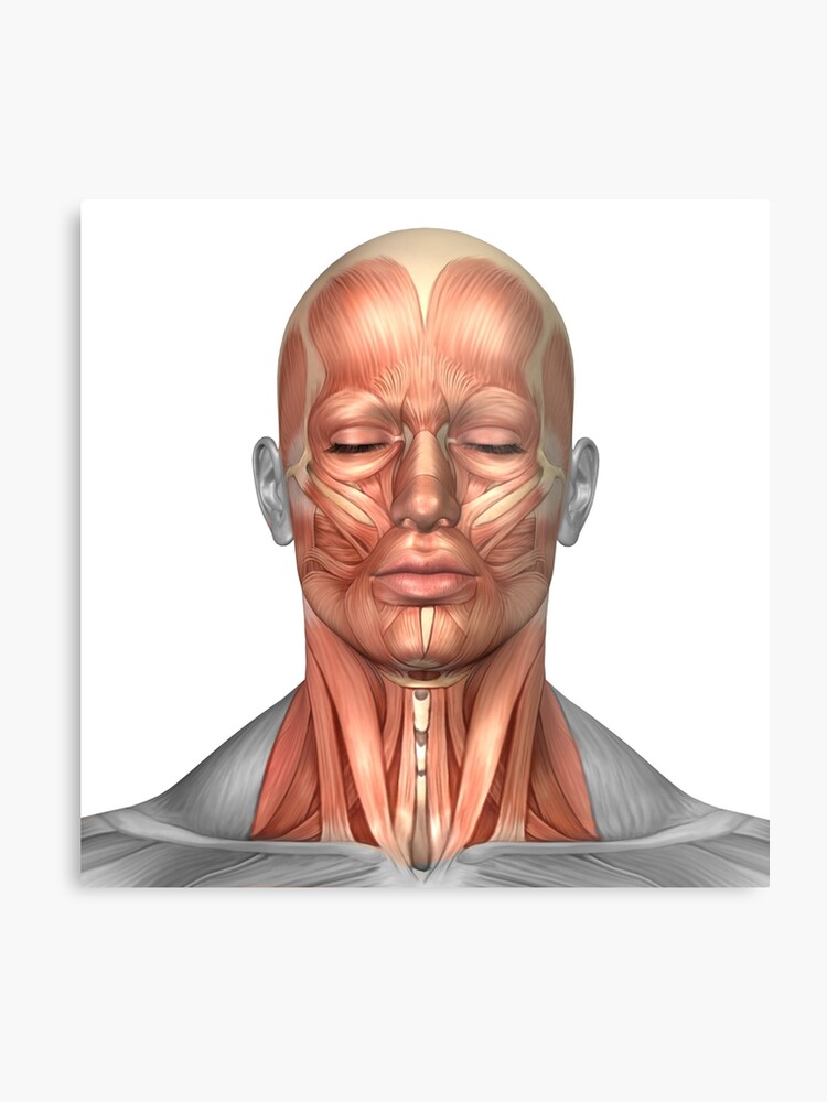 Anatomy Of Human Face And Neck Muscles Front View Metal Print
 Clip Art Neck Muscles Stock Illustration Gg80901812 Gograph
 Anatomy Of The Spine And Back
 Chapter 25 Overview Of The Neck The Big Picture Gross
 Axial Muscles Of The Head Neck And Back Anatomy And
 Cervicogenic Headache Causes And Risk Factors
 Vector Illustration Of Neck Muscles Anatomy
Muscular Anatomy And Soft Tissue Headandcervicalspine
 Neck Atlas Of Anatomy
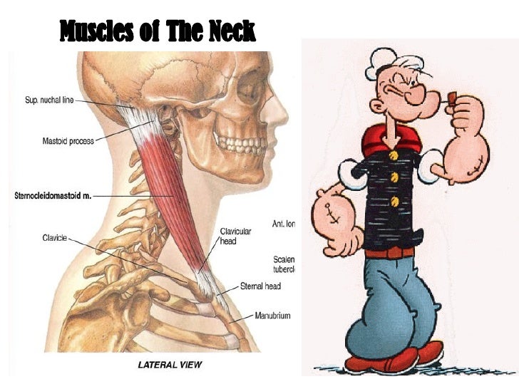 Muscles Of The Neck 1
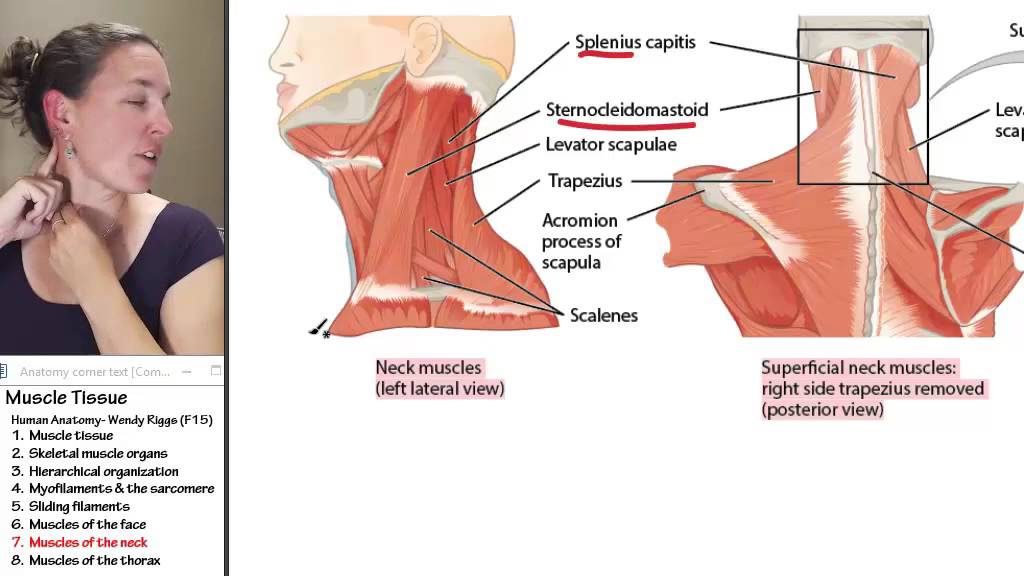 Muscle 7 Muscles Of The Neck
 Muscles Of The Head And Neck Anatomy Pictures And Information
 Yoga Anatomy Forward Head Posture Part 1 Yogauonline
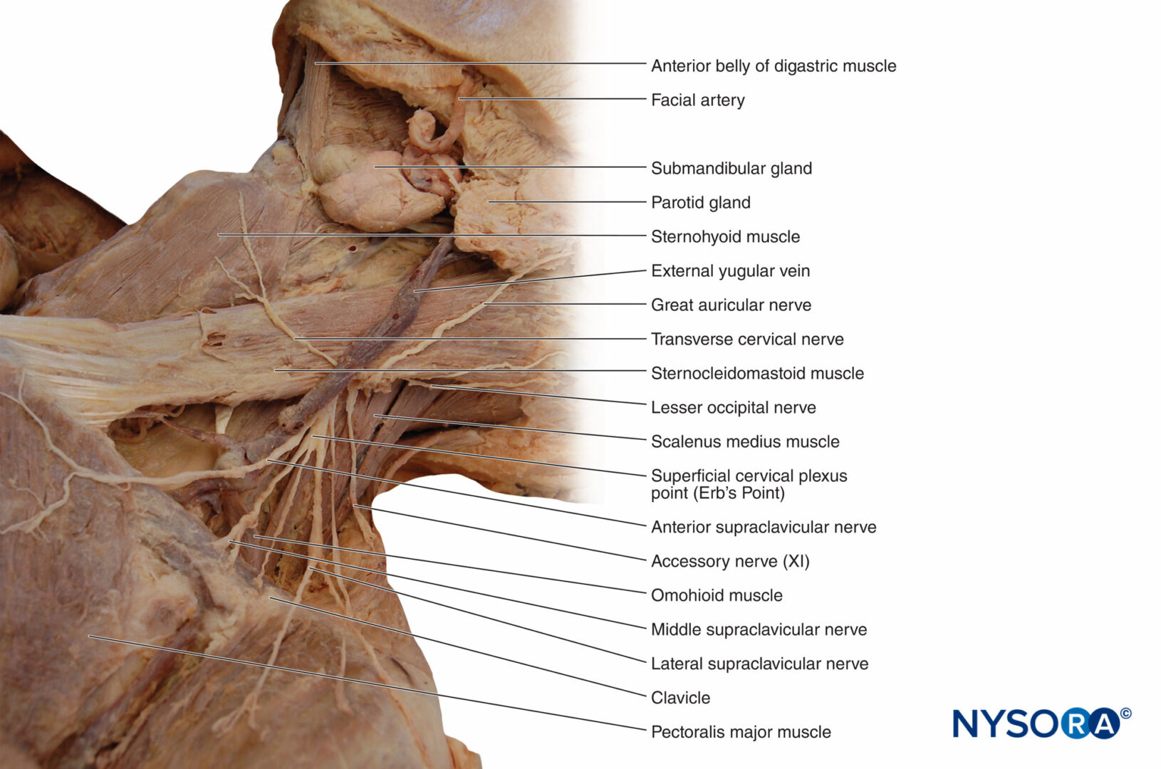 Functional Regional Anesthesia Anatomy Nysora

 Head And Neck Muscles Boundless Anatomy And Physiology
 Crossfit Cervical Muscles Part 2

Belum ada Komentar untuk "Anatomy Of Cervical Muscles"
Posting Komentar