Anatomy Of Heart Structure
Chambers of the heart. About 1 of the cardiac muscle cells in the heart are responsible for forming the conduction system that sets the pace for the rest of the cardiac muscle cells.
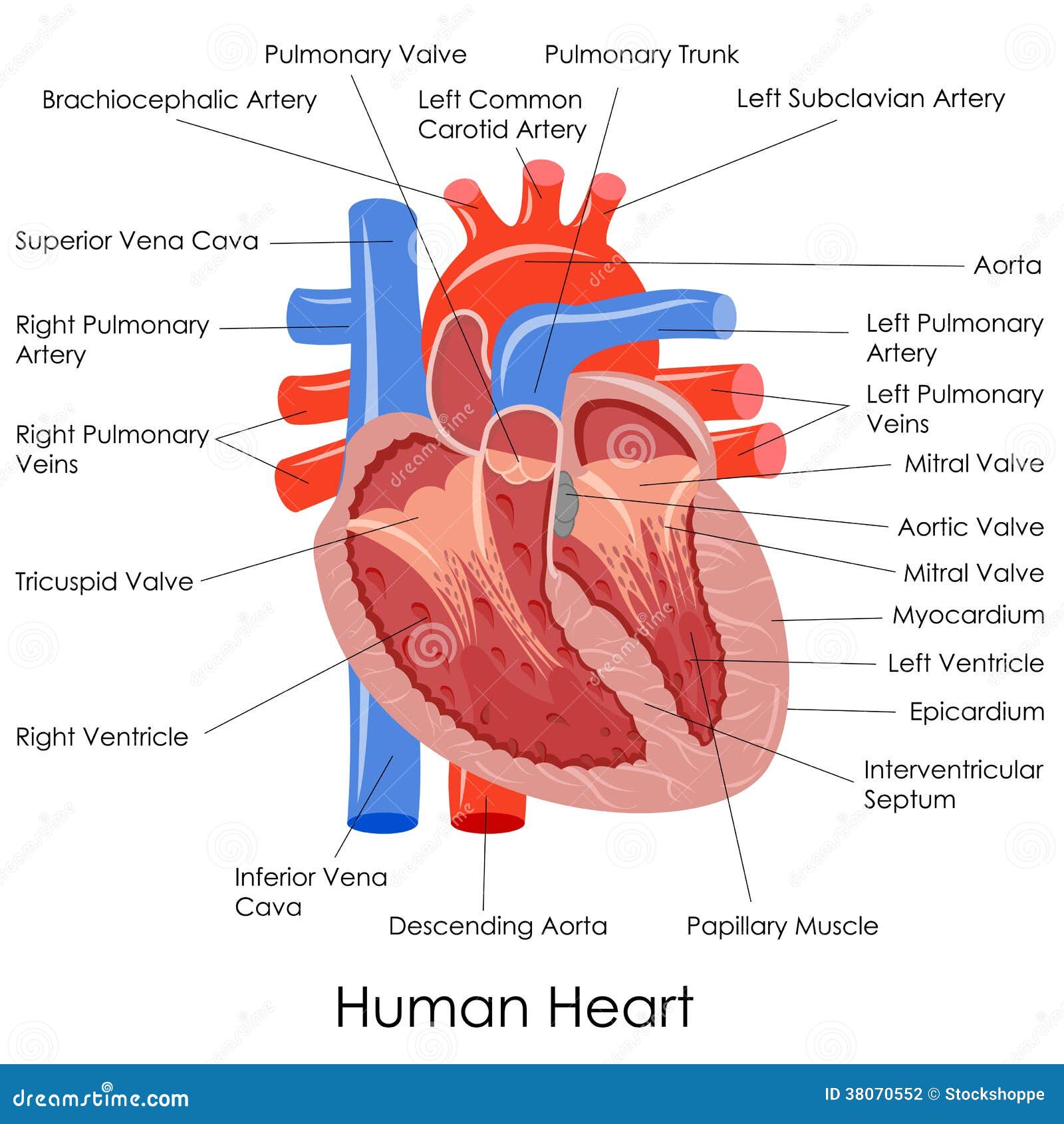 Human Heart Anatomy Stock Vector Illustration Of Anatomy
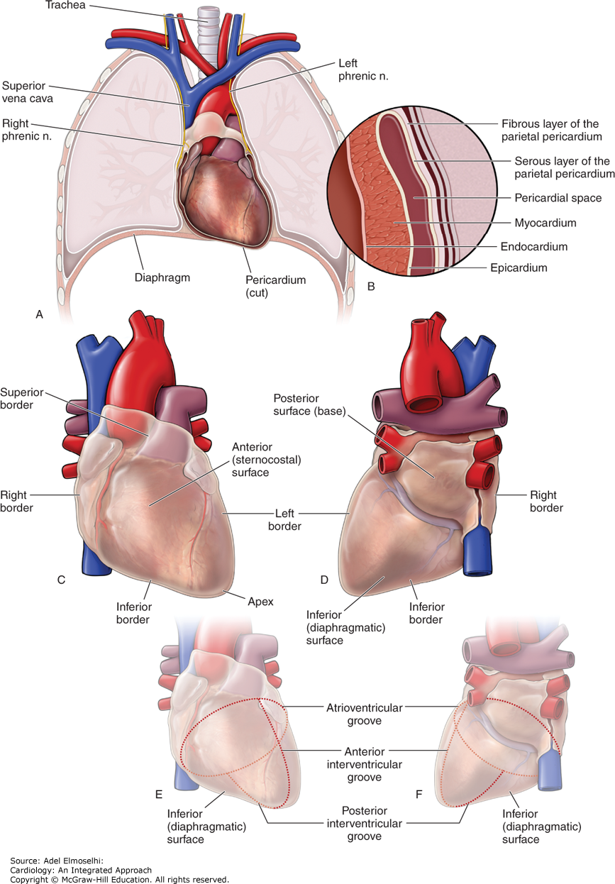
Base posterior diaphragmatic inferior sternocostal anterior and left and right pulmonary surfaces.

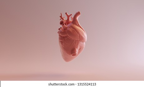
Anatomy of heart structure. The upper two chambers are called the atria. A typical heart is approximately the size of your fist. The anatomy of the heart.
The shape of the heart is similar to a pinecone rather broad at the superior surface and tapering to the apex. 12 cm 5 in in length 8 cm 35 in wide and 6 cm 25 in in thickness. The hearts unique design allows it to accomplish the incredible task of circulating blood through the human body.
Conduction system of the heart. This 3d medical animation depicts the anatomy of the heart. Shape and size of the heart.
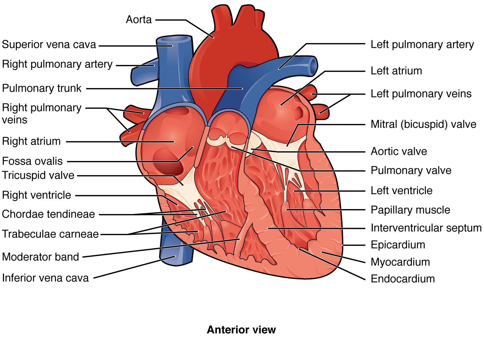
Right left superior and inferior. The heart is a hollow organ with four flexible and resilient inner chambers. Here we will review its essential components and how and why blood passes through them.
This amazing muscle produces electrical impulses that cause the heart to contract. Singly one is called an atrium from the latin word meaning entrance the lower chambers are called the ventricles from the latin word for belly the heart is also divided into two sidesleft and right. Layers of the heart wall.
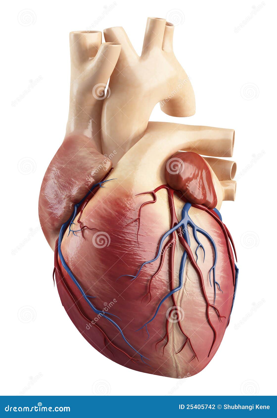
The heart has three layers of tissue each of which serve a slightly different purpose. The heart pumps blood through the network of arteries and veins called the cardiovascular system. The heart has five surfaces.
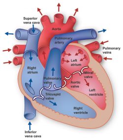
The heart has four chambers. The heart is situated within the chest cavity and surrounded by a fluid filled sac called the pericardium. The heart is a muscular organ about the size of a fist located just behind and slightly left of the breastbone.
The chambers of the heart are shown including the right atrium left atrium right ventricle and left ventricle. Heart anatomy inner chambers. The right atrium receives blood from the veins and pumps it to the right ventricle.
The heart is able to both set its own rhythm and to conduct the signals necessary to maintain and coordinate this rhythm throughout its structures. The right margin is the small section of the right atrium that extends between the superior and inferior vena cava. It is divided by a partition or septum into two halves and the halves are in turn divided into four chambers.
It also has several margins.
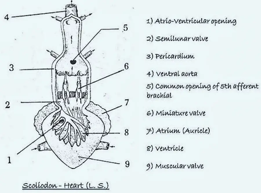 Comparative Anatomy Heart Structure Of Frog And Fish
 Original Silver Anatomical Heart Locket
 Heart Anatomy Anatomy And Physiology
 Heart Anatomy Review
 Heart Structure Medical And Anatomy Poster
 Anatomical Heart
 Cow Heart Structure Reading Industrial Wiring Diagrams

 Wear Your Heart On Your Sleeve With These Anatomical Heart
 Anatomical Structure Of The Heart Cardiology An
Heart Wikipedia
 Human Heart Shirts Anatomical Heart Anatomical Shirts Anatomy Shirts Bc19
 Amazon Com Lxx Human Anatomical Heart Model Life Size

 Heart Structure Anatomy Physiology Wikivet English
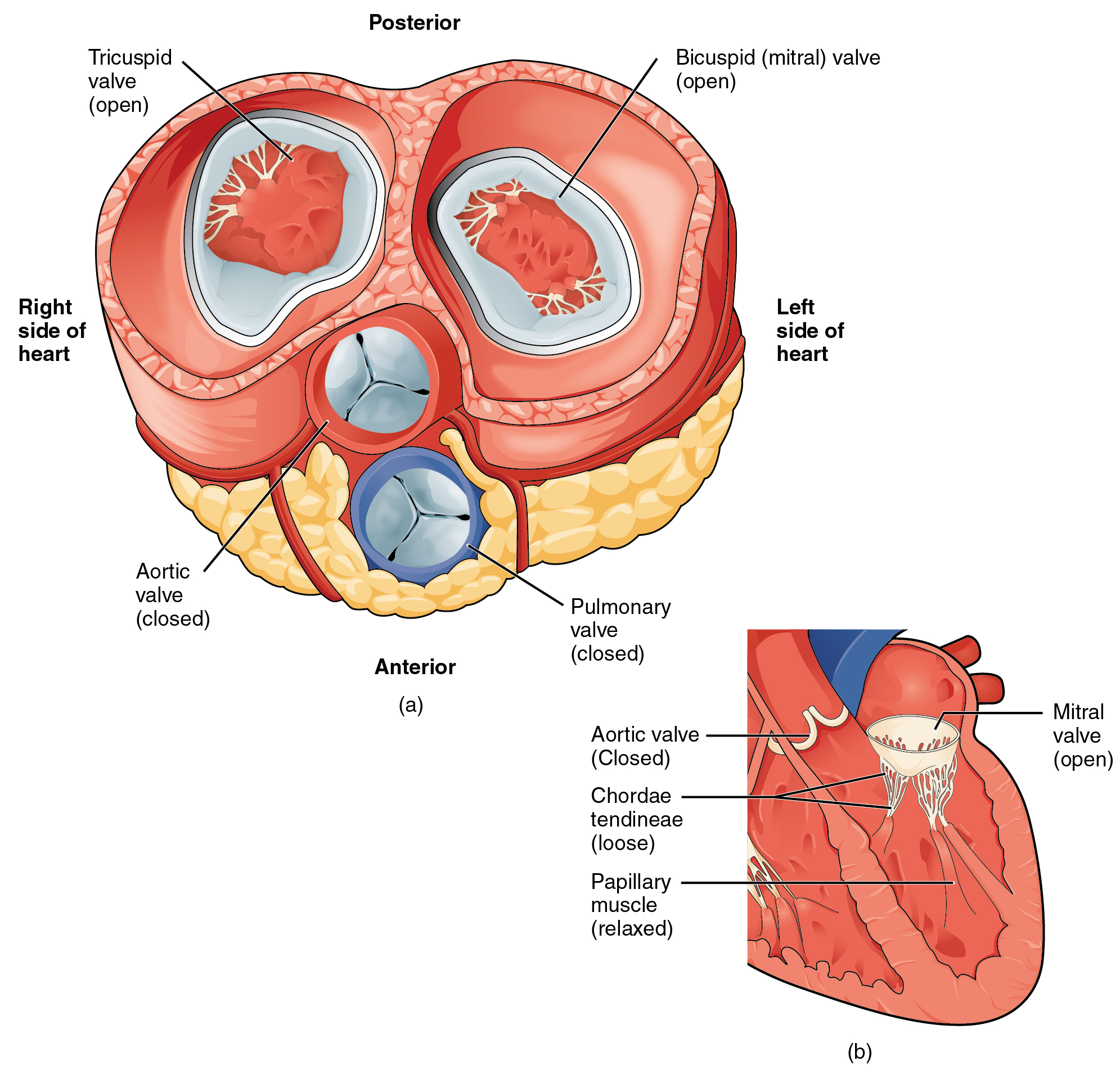
 Cardiac Valves Stock Illustrations Images Vectors
 Cow Heart Structure Reading Industrial Wiring Diagrams
 19 1 Heart Anatomy Anatomy And Physiology
 Realistic Vector Illustration Human Healthy Heart Stock
 Front View Of The Anatomy Of Heart Interior Struct Stock
 Heart Structure Anatomy Physiology Wikivet English
 Anatomy Clipart Real Heart Anatomical Heart Vector Png

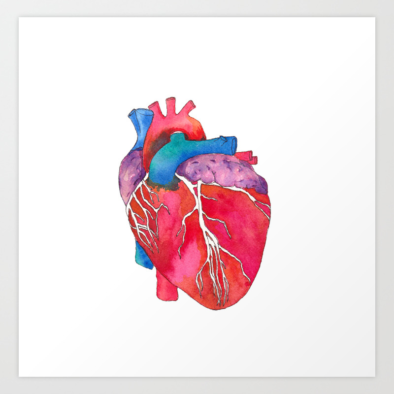 Anatomical Heart Art Print
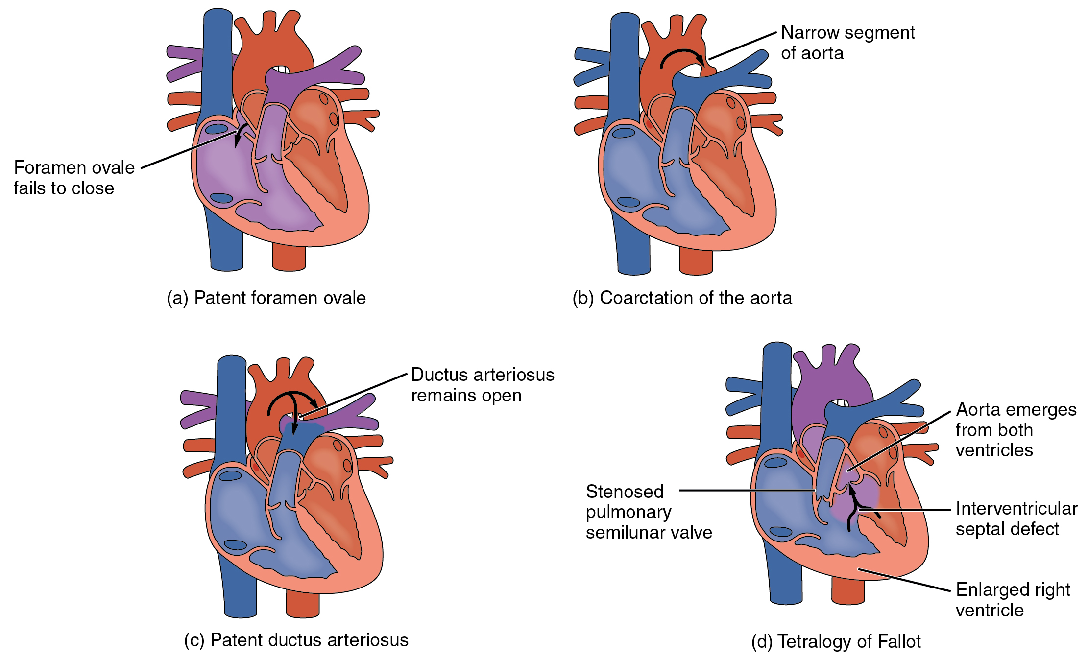 Heart Anatomy Anatomy And Physiology
 Heart Information Center Heart Anatomy Texas Heart Institute
Belum ada Komentar untuk "Anatomy Of Heart Structure"
Posting Komentar